Skip To Main Content
Lilly
Menu closed
Lilly
  • Account Login / Register
  • Medical Home
  • Medical Education
      • All Therapeutic Areas
        • Alopecia Areata
        • Atopic Dermatitis
        • Dermatology
        • Psoriasis
        • Diabetes
        • Hypoglycemia
        • Inflammatory Bowel Disease
        • Crohn's Disease
        • Ulcerative Colitis
        • Migraine
        • Alzheimer's Disease
        • Cognitive Health
        • Obesity
        • Obstructive Sleep Apnea
        • Breast Cancer - Early
        • Breast Cancer - Metastatic
        • Hematological Malignancy
        • Non-Small Cell Lung Cancer
        • Oncology Precision Medicine
        • Thyroid Cancer
        • Axial Spondyloarthritis
        • Psoriatic Arthritis
        • Resources
      • All Therapeutic Areas
        • Alopecia Areata
        • Atopic Dermatitis
        • Psoriasis
        • Diabetes
        • Hypoglycemia
        • Inflammatory Bowel Disease
        • Cluster Headache
        • Migraine
        • Alzheimer's Disease
        • Cognitive Health
        • Obesity
        • Obstructive Sleep Apnea
        • Breast Cancer - Metastatic
        • Gastrointestinal Cancer
        • Hematological Malignancy
        • Oncology
        • Osteoporosis
        • Rheumatoid Arthritis
        • Resources
  • Independent Medical Education
    • Cardiovascular
      • Alopecia Areata
      • Atopic Dermatitis
      • Psoriasis
      • Diabetes
      • Crohn's Disease
      • Ulcerative Colitis
      • Migraine
      • Alzheimer's Disease
      • Obesity
      • Obstructive Sleep Apnea
      • Breast Cancer
      • Gastrointestinal Cancer
      • Hematological Malignancy
      • Non-Small Cell Lung Cancer
      • Oncology
      • Oncology Precision Medicine
      • Prostate Cancer
      • Axial Spondyloarthritis
      • Psoriatic Arthritis
  • Chat Contact Us
Lilly

You are now leaving the Lilly Medical Education website

The link you clicked on will take you to a site maintained by a third party, which is solely responsible for its content. Lilly USA, LLC does not control, influence, or endorse this site, and the opinions, claims, or comments expressed on this site should not be attributed to Lilly USA, LLC. Lilly USA, LLC is not responsible for the privacy policy of any third-party websites. We encourage you to read the privacy policy of every website you visit.
Click "Continue" to proceed or "Return" to return to Lilly Medical Education.

Implementing Biomarker-Informed Cancer Care in NSCLC

Loading icon

Non-Small Cell Lung Cancer

Non-Small Cell Lung Cancer

Most cases (47.6%) of non–small cell lung cancer (NSCLC) cases are advanced or metastatic at diagnosis,1 and 54% of early-stage cases will eventually progress to stage IV.2

Patients with advanced or metastatic NSCLC may be eligible to receive targeted therapy and should have comprehensive genomic profiling performed to test for actionable biomarkers.3

However, less than 40% of eligible patients with NSCLC receive targeted therapy.4 Practice gaps in the NSCLC treatment journey contribute to this patient loss.4 Implementing strategic solutions to standardize and optimize protocols in key workflow areas may help improve overall patient access to targeted therapies. Click below to learn more about each step of the NSCLC treatment journey.

  • Patient Identification

  • Sample Collection

  • Tissue Stewardship

  • Selecting a Molecular Test

  • Multidisciplinary Communication

  • Treatment Decision

  • Managing Disease Progression


Patient Identification

Identifying patients who will benefit from precision medicine and selecting the right first-line therapy is essential to optimizing patient outcomes in NSCLC.5,6 Inappropriate first-line therapies based on incomplete testing are unlikely to be effective and can lead to increased toxicity with subsequent targeted treatments. To optimize selection of the most appropriate first-line therapy, comprehensive molecular testing, in combination with conservative use of IHC, should be performed at diagnosis before initiating treatment in all patients with advanced or metastatic NSCLC.


Actionable Genomic Alterations in NSCLC

More than half of all patients with NSCLC harbor potentially actionable genomic alterations; however, many of these individual alterations occur with relatively low frequency.7 Thus, identifying patients who will benefit from precision medicine often involves detecting relatively rare gene alterations. Successfully doing so allows for targeting of actionable alterations with precision medicine, which potentially improves survival in patients with NSCLC.5,6

The use of targeted therapy has been associated with a more favorable outcome in advanced NSCLC, with a 31% reduction in risk of death and improved survival duration that was approximately 1.5-fold longer compared with patients with an identified mutational driver but did not receive targeted therapy.8 Comprehensive testing is recommended to identify potentially actionable biomarkers and optimize choice of therapy in patients with NSCLC.9

Frequency of Genomic Alterations in NSCLC Adenocarcinoma1

Frequency of Genomic Alterations in NSCLC Adenocarcinoma
chevron-filled-down View image description


KRAS Pathway10,11

  • RAS is a small, membrane-localized GTPase that integrates a number of proliferative signals to establish a tumorigenic cellular circuit when aberrantly activated10
  • RAS signaling is activated by a number of cellular receptors including receptor tyrosine kinases, G protein-coupled receptors, and integrin family members10
  • These signaling cascades initiate RAS activation through assembly of several scaffolding proteins that mediate conversion of RAS from an inactive GDP-bound form to an active GTP-bound state10
  • Following activation, RAS activates the mitogen-activated protein kinase, PI3K-AKT, and Rac-Rho signaling networks10
  • This leads to a variety of functions that promote cancer development, including oncogenic transcription, cell cycle progression, cellular survival, cell growth and metabolism, and cell motility and migration10
  • KRAS inhibitors selectively target a mutant form of KRAS and limit its activation by favoring binding to GDP, blocking its downstream signaling through RAF, and by selectively reducing the frequency of the active, GTP-bound KRAS10

KRAS signaling pathway

KRAS Exons12-17


KRAS exon map with sensitizing and resistance mutations


Sensitizing mutations12

  • G12C
    • G12X mutations comprise 86% of KRAS mutations
    • G12C mutations comprise 47% of G12 mutations


Resistance mutations13

  • KRAS inhibitor binding pocket: Y96C, R68S, H95D/Q/R
  • G12D/R/V/W activating mutation not targetable by G12C inhibitor
  • G12C allele amplification
  • Others: G13D, Q61H


Appropriate testing methodologies14,15

  • NGS (point mutations, amplification)
  • PCR-based methods (point mutations)


EGFR Pathway10,11,18

  • EGFR is a transmembrane receptor protein activated by the binding of specific ligands including epidermal growth factor and transforming growth factor-alpha11,18
  • EGFR is activated either as a homo- or heterodimer resulting in regulation of multiple pathways11,18
  • In particular, the RAS/RAF/MAPK, PI3K/AKT/mTOR, and JAK/STAT pathways downstream of EGFR play integral roles in cell migration, proliferation, and survival, respectively10,11
  • Anti-EGFR antibodies are targeted to the external ligand-binding domains, while the small molecule inhibitors or tyrosine kinase inhibitors target their cytoplasmic kinase domains11

EGFR signaling pathway

EGFR Exons14,16,18-20


EGFR exon map with sensitizing and resistance mutations


Sensitizing alterations16,18

  • Exon 18 mutations (G719C/S/A) ~4%
  • Exon 19 deletions ~44%
  • Exon 20 insertions/in-frame duplications ~5%
  • Exon 20 mutations (V765A, T783A) <1%
  • Exon 21 mutations (L858R) ~41%
  • Other Missense mutations ~6%


Resistance mutations14,18

  • T790M ~50%
  • Exon 20 insertions (relatively rare)


  • Appropriate testing methodologies14,19

  • NGS (point mutations, indels, amplification)
  • PCR-based methods (point mutations)


  • ALK Pathway10,11,21,22

    • ALK is a member of the insulin receptor superfamily of cellular transmembrane receptors possessing intrinsic tyrosine kinase activity21
    • Wild-type ALK is activated by ligand-dependent (pleiotrophin and midkine) receptor dimerization and autophosphorylation21
    • Activated ALK triggers downstream signaling pathways, including PI3K-AKT, JAK-STAT and MAPK pathways, which control a variety of cellular processes22
    • Ligand-independent ALK activation occurs through fusion protein formation and activating ALK point mutations21
    • Ligand-dependent ALK activation occurs through ALK amplification21
    • Selective ALK inhibitors allow for the potent suppression of cell growth in tumors expressing ALK fusion proteins22

    ALK signaling pathway

    ALK Exons14,23-27


    ALK exon map with sensitizing and resistance mutations


    Sensitizing alterations23

    • Known fusions
      • >85% EML4-ALK23,24
      • Other fusion partners identified to date: KIF5B, KLC1, and TFG23,25
      • Novel ALK fusions also possible23
    • ALK amplification/overexpression25


    Resistance mutations23,25

    • L1196M (gatekeeper mutation)
    • G1202R
    • S1206Y
    • Other resistance mutations: C1156Y, G1269A, F1174L, L1198P, and D1203N
    • ALK copy number gain


    Appropriate testing methodologies14,27

    • NGS (point mutations, fusions, amplification)
    • FISH (amplification, fusions)
    • IHC (overexpression)


    BRAF Pathway11,28,29

    • BRAF is 1 of 3 isoforms of RAF (ARAF, BRAF, and CRAF)28
    • It is the upstream kinase of MEK, and the direct effector of RAS, which delineates the whole RAS/RAF/MEK/ERK signaling pathway29
    • BRAF inhibitors target oncogenic alterations on BRAF that aberrantly activate the RAS/RAF/MEK/ERK signaling pathway29

    BRAF signaling pathway

    BRAF Exons14,30-34


    BRAF exon map with sensitizing and resistance mutations


    Sensitizing mutations30

    • V600E ~50% of BRAF mutations
    • V600K/D/M ~1-2%


    Resistance mutations (mostly off target)31

    • Rare gatekeeper mutations L505H and L514V


    Appropriate testing methodologies14,32

    • NGS
    • PCR-based methods
    • IHC (overexpression)


    MET Pathway10,11,35,36

    • MET is a glycoprotein transmembrane receptor tyrosine kinase35
    • MET activation occurs when HGF ligand binds to the MET receptor, leading to homodimerization and phosphorylation of intracellular tyrosine residues35
    • This in turn leads to activation of the downstream RAS/ERK/MAPK, PI3K/AKT and STAT signaling pathways35
    • Finally resulting in cell proliferation, survival, migration, motility, invasion, angiogenesis, and the epithelial-to-mesenchymal transition, depending on the cell type35
    • MET gene dysregulations in lung cancer can occur in multiple ways including gene mutation, amplification, rearrangement, and protein overexpression35
    • Several agents with different mechanism of actions to target MET are available, such as multikinase and selective MET inhibitors35

    MET signaling pathway

    MET Exons14,35,37-40


    MET exon map with sensitizing and resistance mutations


    Sensitizing alterations35,37

    • MET amplification/overexpression ~5%
    • MET exon 14 alterations at splice sites (SNVs, indels) ~4%
      • Leads to exon 14 skipping/deletion
    • Point mutations (<1%)35,37
      • Juxtamembrane domain: Y1003X and D1010H
      • Catalytic domain: D1228H, Y1235D, M1250T


    Resistance mutations38

    • MET exon 14 mutant allele amplification
    • Secondary kinase domain mutations: H1094Y, G1163R, L1195V, D1228H/N, and Y1230C/H/S


    Appropriate testing methodologies14,39

    • NGS (fusions, exon 14 skipping, point mutations, amplification)
    • PCR-based methods (point mutations)
    • FISH (amplification, fusions)
    • IHC (overexpression)


    ROS1 Pathway10,11,41

    • ROS1 is an orphan transmembrane tyrosine kinase receptor41
    • Dysregulated ROS1 may occur as a result of ROS1 gene fusion, overexpression, or mutation41
    • Aberrant ROS1 kinase activity leads to activated downstream signaling of several oncogenic pathways including PI3K/AKT/mTOR and RAS-MAPK/ERK pathways41
    • Given the high homology (49% amino acid homology within the kinase domain and 77% identity at the ATP-binding site) in the kinase domains of ROS1 and ALK, ALK inhibitors are efficacious against ROS1-positive cell lines and tumors41

    ROS1 signaling pathway

    ROS1 Exons14,41-44


    ROS1 exon map with sensitizing and resistance mutations


    Sensitizing alterations41,42

    • Known fusions
      • CD74 and SDC4 are most common
      • Other fusion partners identified to date: SLC34A2, EZR, LRIG3, TPM3, and FIG
    • Novel ROS1 fusions also possible


    Resistance mutations (~50% on target)42

    • G2032R, D2033N, L2026M, L2000V, L2086F, 1951R, S1986F/Y


    Appropriate testing methodologies14,43

    • NGS (fusions)
    • FISH (fusions)
    • FISH (amplification, fusions)
    • IHC (overexpression – screening only)


    HER2 Pathway45,46

    • HER2, encoded by ERBB2, is a transmembrane glycoprotein receptor with intracellular tyrosine kinase activity belonging to the EGFR family (EGFR/HER1, HER2/3/4)45,46
    • HER2 does not have a known soluble ligand and downstream signaling is triggered by dimerization with other ligand-bound HER family members45
    • Activation of this receptor tyrosine kinase family triggers a cascade of subcellular signal transduction pathways controlling cell growth, differentiation, and motility45
    • Alterations (amplification, mutations) in the HER2 gene result in hyperactivation following increased homo- or heterodimerization and autophosphorylation, which triggers multiple signaling pathways resulting in uncontrolled cell proliferation45
    • Several methods are available to target HER2 alterations45:
      • Small molecule TKIs
      • Anti-HER2 antibodies
      • Antibody drug conjugates

    HER2 signaling pathway

    HER2 Exons14, 46-48


    HER2 exon map with sensitizing and resistance mutations


    Sensitizing alterations (most frequent listed)46

    • HER2 activating mutations ~2-4%
      • S310F/Y, R678Q, L755P/S, D769Y, V777L/M, V842I
      • Exon 20 insertions, duplications (most common)
    • HER2 amplification ~10-20%
    • HER2 overexpression ~2.4-38%


    Resistance mutations (data still emerging)46

    • L755S, V777L, D769Y, V842I, K753E, I655V


    Appropriate testing methodologies14,47

    • NGS (point mutations, amplification)
    • PCR-based methods (point mutations)
    • FISH (amplification)
    • IHC (overexpression)


    RET Pathway10,11,49-51

    • RET is a transmembrane glycoprotein receptor tyrosine kinase encoded by the proto-oncogene RET49,50
    • GDNF ligands bind to GDNF family receptor α (GFRα) co receptors, which mediate RET homodimerization, resulting in trans autophosphorylation of tyrosine residues within the RET intracellular domains, recruitment of key signaling adaptors, and activation of several signal transduction cascades involved in cellular proliferation, including the MAPK, PI3K, JAK-STAT, PKA, and PKC pathways49,50
    • Oncogenic activation of RET occurs by 2 primary mechanisms49,50:
      • Chromosomal rearrangements produce hybrid proteins that fuse the RET kinase domain with a partner protein that often contains a dimerization domain, resulting in ligand-independent dimerization and constitutive activation of the kinase
      • Mutations that directly or indirectly activate the kinase
    • RET inhibitors act as ATP mimetics to displace ATP, blocking the signaling activity of the fusion oncogenes and causing either apoptosis or proliferation inhibition50

    RET signaling pathway

    RET Exons14,52-56


    RET exon map with sensitizing and resistance mutations


    Sensitizing alterations52,53

    • Known fusions
      • KIF5B-RET and CCDC6-RET are most common
      • Other fusion partners identified to date: NCOA4 and TRIM33
      • Novel RET fusions also possible


    Resistance mutations (data still emerging, ~10% on target)54

    • G810C/S solvent-front mutation
    • V804M gatekeeper mutation
    • Y806C


    Appropriate testing methodologies14,55

    • NGS (fusions, point mutations)
    • PCR-based methods (point mutations)
    • FISH (fusions)


    NTRK1/2/3 Pathway51,57

    • NTRK1/2/3 encode TRKA/B/C, respectively, of the TRK family. TRK proteins contain an extracellular ligand-combining domain, a transmembrane domain, and an intracellular tyrosine kinase domain51,57
    • TRKA/B/C bind to nerve growth factor, brain-derived growth factor, and NT-3, respectively, and promote cell proliferation and survival through downstream MAPK/ERK, PLCγ/PKC, and PI3K/AKT signaling51,57
    • Dysregulation of this pathway and subsequent tumorigenesis occurs through ligand-independent constitutive activation TRKA/B/C51,57
    • This constitutive activation is generally the result of genomic rearrangement between NTRK1/2/3 and a fusion partner gene51,57
    • Most functional NTRK fusion proteins lack the extracellular ligand-combining domain of TRK; instead, the partner gene is expressed in a homogenous dimer, which induces ligand-independent activation of the tyrosine kinase domain and upregulates downstream cancer-associated pathways51,57

    NTRK1/2/3 signaling pathway

    NTRK Exons51, 58-63


    NTRK1/2/3 exon map with sensitizing and resistance mutations


    Sensitizing alterations58

    • Known fusions
      • TPM3-NTRK1 and ETV6-NTRK3 are most common
      • Other fusion partners found in lung adenocarcinomas: CD74, MPRIP, TRIM24, and VANGL2
      • Novel NTRK fusions also possible


    Resistance mutations (solvent-front/gatekeeper)51

    • NTRK1 F589L, G595R, G667C/S
    • NTRK2 G639R
    • NTRK3 F617L, G623E/R, G696A


    Appropriate testing methodologies59

    • NGS (fusions, point mutations)
    • FISH (fusions)
    • IHC (overexpression – screening only)


    Immune Checkpoint Inhibitors in NSCLC
    antibody

    PD-1 and PD-L1 Testing

    PD-L1 expression is the currently available biomarker to assess whether patients are responsive to PD-1 or PD-L1 inhibitors.64,65 However, it is important to obtain molecular test results for actionable genomic biomarkers before administering immune checkpoint inhibitors (ICIs) in the first line, because when given alone, ICIs have low efficacy in patients with oncogenic-driven metastatic NSCLC.9,66 Multiple anti-PD-L1 IHC assays have been developed for different ICIs. Definition of a positive or negative PD-L1 test result depends on the individual antibody, clone, and platform, which may be unique to each ICI.64,65
    mismatched puzzle pieces

    Microsatellite Instability and DNA Mismatch Repair

    DNA MMR pathways play a key role in identifying and repairing mismatched nucleotides that arise during genetic recombination or because of damage caused by extrinsic physical or chemical insults. If one or more MMR proteins are not expressed or are dysfunctional, the tissue is considered dMMR.67 Inactivation of MMR genes can result in microsatellite instability.67 ICI therapy has been shown to be effective in dMMR/MSI-high GI and endometrial tumors, but there is limited evidence in NSCLC.68
    mutated DNA strand

    Tumor Mutational Burden

    TMB is the total number of somatic mutations in a defined region of a tumor genome and varies according to tumor type as well as among patients.69 Some clinical data demonstrate that tumors with high TMB are more likely to achieve clinical benefit from treatment with ICIs.70 However, several other trials have shown that high TMB levels do not correlate with PD-L1 expression levels in patients with NSCLC.70 Moreover, some patients with low TMB levels respond to immunotherapy, and others with high levels do not respond to immunotherapy.70

    Sample Collection


    Collection Technique

    Collection technique affects diagnostic yield and downstream success of genomic testing (molecular yield). Identifying the reason for biopsy can help choose the best method to optimize yield.71-73

    Thoughtful selection of the appropriate biopsy approach (eg, technique, needle gauge) for specimen collection can ensure sufficient material is available to render a diagnosis and provide comprehensive biomarker testing from a single procedure.74

    If initial biopsy reveals lung adenocarcinoma, but limited tissue remains after diagnostic workup75:

    • Communicate presence of limited testing material to the ordering provider
      • Prioritize testing for targets most likely to be identified and/or smaller panels that are less likely to be QNS
      • Consider ordering a liquid biopsy (ctDNA assay)
      • Consider repeat biopsy, and communicate “molecular priority” protocol for known diagnosis
      • Consider testing alternative specimens (see “Tissue Stewardship”)

    Approximate Diagnostic Yield of Tissue Collection Techniques71-73

    Approximate Diagnostic Yield of Tissue Collection Techniques
    chevron-filled-down View image description

    ROSE has the potential to positively impact outcomes.76-80

    ROSE has the potential to positively impact outcomes
    chevron-filled-down View image description
    Rapid OnSite Evaluation (ROSE) has the potential to positively impact outcomes by increasing diagnostic accuracy and yield. Cytopathologic diagnostic assessment of individual biopsy passes performed during a biopsy procedure can help distinguish between diagnostic adequacy and ancillary molecular testing adequacy.76-80

    Each molecular laboratory will have a minimal amount and concentration of tumor cells required for accurate detection of molecular alterations, based on the specific tumor enrichment protocols available and assay platforms used for testing.81 Most NGS platforms require a sample size of ≥25 mm² tumor surface area and ≥20% viable tumor nuclei per sample.82,

    Tissue blocks that are adequate for molecular testing should be tracked and by flagging in the report for the tissue navigator and/or lab technician.84

    Tissue Stewardship

    While resection specimens have abundant tissue available for testing, they are available only in a small subset of patients with NSCLC.81 Thus, it is necessary to consider other types of specimens that might require testing.81 The majority of patients with lung cancer are diagnosed after examination of a small biopsy or cytology specimen of the primary lesion or a suspected metastasis.81 It has been well documented that small biopsy specimens, cytology cell blocks, and cytology touch imprints or aspirate slides are suitable for molecular testing.81


    Impact of Acquisition Method on Sequencing Success85

    Impact of Acquisition Method on Sequencing Success Impact of Acquisition Method on Sequencing Success
    chevron-filled-down View image description
    These specimens should be handled prudently with as much material as possible preserved for molecular testing, keeping in mind that they may be the only specimen available.81,84,86 A limited number of immunohistochemical preparations (rather than a “shotgun” panel) is preferable to confirm a pulmonary origin and identify adenocarcinoma.81,84,86 Where possible, limit IHC to PD-L1 status (for fist-line immunotherapy), 1 adenocarcinoma marker, and 1 squamous carcinoma marker.81,84,86

    Implementing standardized tissue-specific procedures and optimizing pre-analytic conditions by processing samples according to lab specifications via a preferred vendor may help reduce quantity not sufficient (QNS) rates and downstream sequencing failure.85,87

    Standardized Tissue Handling
    chevron-filled-down View image description
    Specific methods that can be used to preserve tissue include cutting 15-20 unstained slides up front (which may provide enough slides for molecular testing and leave several unstained slides available if immunostains are needed).81 Although it requires additional time and effort in the histologic processing laboratory, all tissue cut from the block should be picked up onto glass slides (no sections should be discarded from the water bath).81

    Selecting a Molecular Test


    Next-Generation Sequencing Approach

    Although a single gene test (SGT) may have a shorter turnaround time than NGS, NGS is faster than sequential testing with SGTs.88 Upfront NGS testing in metastatic NSCLC is associated with reduced cost and time-to-results for commercial and CMS payers.89

    Additionally, with a sequential single-gene approach, tumor tissue may be insufficient, and some biomarkers may come back without results, which may impact treatment strategy and patient outcomes.86

    Single Gene Test vs Comprehensive Profiling86

    Single Gene Test vs Comprehensive Profiling
    chevron-filled-down View image description
    Comprehensive tumor profiling can be achieved with NGS-based testing. All the recommended biomarkers for NSCLC can be analyzed by broad molecular profiling with NGS sequencing of tumor tissue or circulating tumor DNA.90

    Molecular Testing Options to Identify Targetable, Sensitizing Alterations in NSCLC9,14,47,55,68,90-94

    Molecular Testing Options to Identify Targetable, Sensitizing Alterations in NSCLC
    chevron-filled-down View image description
    Concurrent Tissue and ctDNA Testing89,95

    Use of tissue- and plasma-based (ctDNA) testing are acceptable in the advanced/metastatic NSCLC setting, whether used sequentially or concurrently. Concurrent testing has the potential to reduce TAT if a protracted wait is anticipated for tissue-based results or may also be indicated in cases where there is limited tumor tissue available and QNS is probable. The identification of an actionable driver mutation by either method is sufficient to start therapy. Conversely, the absence of an actionable driver mutation in either assay does warrant the use of a complementary method. Additional advantages and disadvantages to both testing approaches are outlined below.

    Tissue
    Advantages: Pathology information, assessment of DNA and non-DNA biomarkers, PD-L1 assessment

    Disadvantages: Longer TAT, limited tissue quantity/quality, invasive at disease progression, re-biopsy not always feasible, tumor heterogeneity
    ctDNA
    Advantages: Rapid TAT, minimally invasive, repeatable overtime, better capture of tumor heterogeneity and clonal evolution

    Disadvantages: Non-DNA biomarkers are not evaluable, Low tumor fraction, presence of mutations from sites other than the target lesion, most commonly ChIP
    Tissue + ctDNA
    Advantages: All included in ctDNA and tissue testing

    Disadvantages: Potential increased costs

    Diagnostic Algorithm for ctDNA Testing Use in Treatment-naïve Advanced/Metastatic NSCLC89,95

    Diagnostic algorithm for ctDNA testing use in treatment-naïve advanced/metastatic NSCLC
    chevron-filled-down View image description
    The algorithm above is applicable to advanced/metastatic NSCLC. However, there are currently no consensus guidelines in early-stage NSCLC regarding the use of ctDNA testing. When available, a tissue sample should be used if molecular testing is required, and this can be either the diagnostic specimen (if sufficient) or the post-resection specimen. If tissue is unavailable, ctDNA can be considered, but the limitations of the assay must be understood prior to ordering.

    Multidisciplinary Communication


    Doctor Jill Kolesar and Doctor Ravneet Thind

    Learn from Drs. Jill Kolesar and Ravneet Thind about how the multidisciplinary communication facilitated by a molecular tumor board can enable biomarker-informed decision-making and optimize therapeutic options for patients.

    Molecular Tumor Boards

    As molecular diagnostic testing is becoming increasingly sophisticated and complex, molecular tumor boards can bring relevant expertise to this rapidly emerging field and are crucial for patient care.

    Recommended treatment choice increased

    >16% increase in recommended treatment choice96,97

    Time to treatment initiation decreased

    ~30% decrease in time to treatment initiation96-98

    Overall patient survival rate increased

    ~40% absolute increase in overall patient survival rate99


    Expanding Molecular Tumor Boards to Community Settings with Drs Jill Kolesar and Ravneet Thind

    QUESTION 02: What are the advantages of an MTB?

    Dr. Kolesar: We implemented a molecular tumor board with the intention of increasing the rates of molecular testing, which we anticipated would decrease turnaround time for testing, increase access to clinical trials, and ultimately, more patients receiving biomarker-informed treatment decisions.

    Dr. Thind: Due to the general cytotoxicity of conventional chemotherapy, focus has now shifted to novel therapeutic targets that can be exploited based on genomic data and molecular tumor board recommendations. So as Jill mentioned previously, molecular tumor boards can increase guideline-concordant testing and decrease turnaround time for molecular testing, which enables biomarker-informed decision-making and optimizes therapeutic options for our patients.

    Dr. Kolesar: Dr. Thind, as you can see in this figure from our case-control study, we showed that patients with non–small cell lung cancer who had their cases reviewed by a molecular tumor board had improved overall survival compared to those who did not, regardless of whether they were treated in an academic setting or a community.

    MTB Coordinators and Patient Navigators
    Academic centers can expand molecular tumor boards to reach community sites by:

    • Hosting virtual meetings
    • Providing a molecular tumor board-dedicated navigator


    Expanding Molecular Tumor Boards to Community Settings with Drs Jill Kolesar and Ravneet Thind

    QUESTION 02: What elements were necessary for extension of the MTB to rural community sites?

    Dr. Kolesar: I think the most important part was really to make it easy for sites to participate. So to do that, we held a virtual meeting, and we had a molecular tumor board coordinator that managed the cases referred from the community sites.

    Our coordinator interacts with clinic staff from the treating physician office to gather up the notes and NGS reports for review and to provide the written recommendations back. They also coordinate the virtual meeting.


    Dr. Thind: Yes, I agree. I think having a molecular tumor board–dedicated navigator has been a game changer. Along with excellent communication and support from the University of Kentucky, with clear and precise explanation of the entire process.

    Dr. Kolesar: I think it was also really helpful to demonstrate the clinical benefit showing that patients with molecular tumor board review had better outcomes.

    And having wide clinical and genomic expertise, with evidence grading were also critical to the success of the molecular tumor board.

    Dedicated molecular tumor board coordinators and patient navigators can help with100:

    • Consolidation of reports and upload into EMR
    • Follow-up of results and communication of findings
    • Facilitation of multidisciplinary discussions

    MTB coordinators help share reports
    chevron-filled-down View image description

    Treatment Decision


    RET-Positive NSCLC Patient Case Studies
    • Test for certain molecular and immune biomarkers in all appropriate patients with NSCLC
    • Have molecular testing results for the actionable oncogenic mutations before starting systemic therapy combined with immunotherapy
    • Treat patients with oncogenic driver mutations with targeted first-line therapy for that oncogene rather than first-line ICIs


    Welcome everybody. My name is Tim Burns, and I am a principal investigator and thoracic medical oncologist at the University of Pittsburgh Medical Center, Hillman Cancer Center. My clinical and translational research interests focus on the development of novel targeted therapy approaches for oncogene-driven lung cancer.
    Today, we’ll be discussing the case of a patient who was referred into my care. The patient was a 73-year-old female with stage IVa non–small cell lung cancer. At diagnosis, her adenocarcinoma of the lung had begun to spread to the lymph nodes, and it caused a right pleural effusion.
    When I met this patient, unfortunately, she had been admitted into the cardiac intensive care unit for florid new onset of congestive heart failure. Now, we’ll review the patient’s treatment history leading up to this point and after she was transferred into my care.
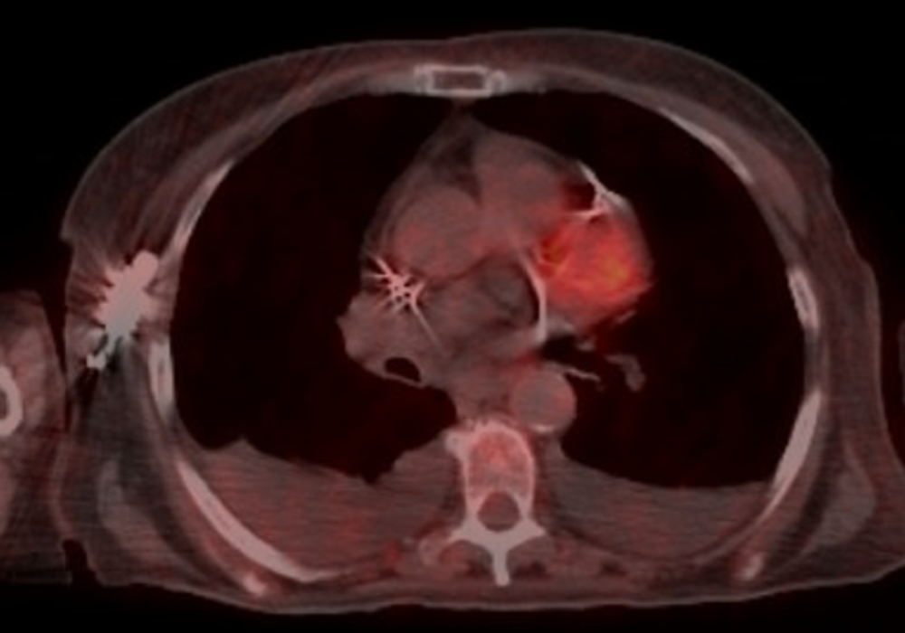
    The patient was a never smoker and denied any drug or alcohol use. She had a history of asthma, and in the 6 months leading up to her diagnosis, she had been treated for 3 occurrences of pneumonia without significant improvement. She eventually presented
    to the emergency room with persistent cough and dyspnea.

    A CT scan at that time found a right middle lobe, right lower lobe mass with mediastinal adenopathy. A subsequent PET/CT scan revealed a PET-avid 7 cm right upper lobe and middle lobe mass-like structure, supraclavicular and large mediastinal lymphadenopathy, and a loculated right pleural effusion.

    The patient underwent a bronchoscopy, EBUS, and biopsy of the mass and lymph nodes at stations 4, 7, and 10. Pathology results were consistent with adenocarcinoma of the lung. Tissue samples were also sent for comprehensive genomic profiling and immunohistochemistry, which revealed a PD-L1 of 70%, a KIF5B-RET fusion, a PIK3CA mutation, and a tumor mutation burden of
    6.9 mutations/Mb.

    So, in this patient, there are several potential targetable molecular biomarkers that we found. First, the patient has a targetable RET fusion in her tumor, and there are first-line targeted therapies with 1 of 2 selective RET TKIs, which has been shown to be superior both in response rate and progression-free survival compared to chemotherapy. And so clearly the choice for this patient would be to treat the RET fusion with a selective RET inhibitor.
    You also see a PIK3CA mutation. In lung cancer, although there are PIK3CA inhibitors that are approved for breast cancer; there are none that are approved in lung cancer. And to date, there really has not been a lot of activity of these inhibitors in lung cancer. In addition, you'll notice there's a high PD-L1 expression here. Well, it's often the case when you have an oncogenic driver that you'll see high levels of PD-L1. And that's actually because a lot of these oncogenic drivers will drive PD-L1 expression. However, in that case, this high expression is unrelated to whether these tumors can respond to immunotherapy, and that should not be used as a way of selecting patients who respond to immunotherapy.
    Furthermore, previous studies have shown that there's almost no benefit to the addition of immunotherapy to chemotherapy in the first-line setting for these
    RET fusion–positive non–small cell lung cancer patients.

    Despite what I just said on the last slide, this patient was initially cared for at an outside institution, and they were given chemoimmunotherapy in the first-line setting. She received 2 cycles of therapy until, unfortunately, she presented to the emergency room, now with right arm pain and swelling and was found to have new arterial thrombosis requiring an embolectomy and also had a DVT. She also had a CT scan at that time, which now revealed worsening right lower lobe opacification, new and enlarged left lobe nodules, and a right pleural effusion, which had increased and would now require a thoracentesis.
    After discharge, she received 1 final cycle of Pembrolizumab alone. Two weeks following this treatment, she was admitted into the cardiac intensive care unit with florid new onset congestive heart failure and required a left ventricular assist device and a pacemaker for now complete heart block. A transthoracic echocardiogram revealed an ejection fraction of only 10%-15%. A right heart catheterization and biopsy confirmed autoimmune myocarditis.
    This patient was aggressively treated with immunosuppressive therapy, and luckily her ejection fraction improved to 40%-45%. A CT scan of the chest during this admission demonstrated a modest response to therapy in the mediastinal adenopathy and right-sided consolidation; however, she had an increase in her right pleural effusion.
    So, at this point we have a patient that's clearly progressing and has had serious side effects from her chemoimmunotherapy. She has required aggressive care for her autoimmune myocarditis, and at this point any further immunotherapy would be absolutely contraindicated. If this patient were to be rechallenged with immunotherapy, it is likely that she would potentially die from her myocarditis if she had another attack.
    Given that chemoimmunotherapy is no longer an option, the question is what should we do next for this patient. And I think this is, as we talked about before, we have 2
    FDA-approved RET-specific inhibitors that we could use for this patient. And I think that would be a reasonable approach.

    After discharge, the patient followed up in our clinic to discuss next steps in her therapy. She was now on anticoagulation for her recent clots and on medication both for her heart failure and steroid-induced diabetes. Despite this, she had been feeling well since discharge, with some rare lightheadedness but no new or concerning symptoms.
    She was started on a selective RET inhibitor, and the patient was followed closely with visits every 2 weeks for side effect monitoring, laboratory work, and EKG monitoring.
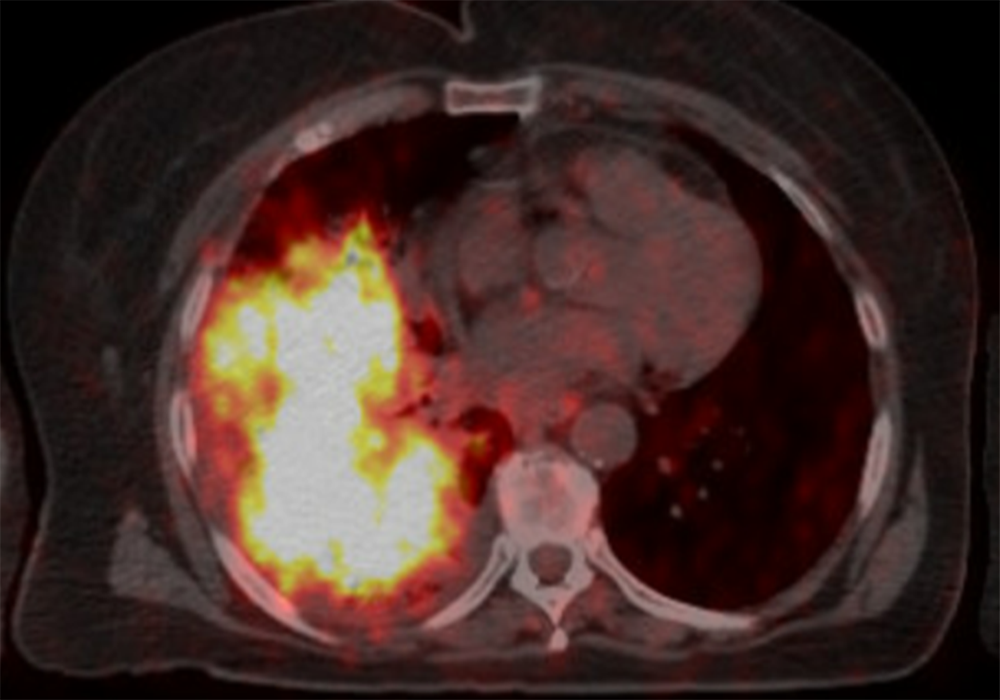
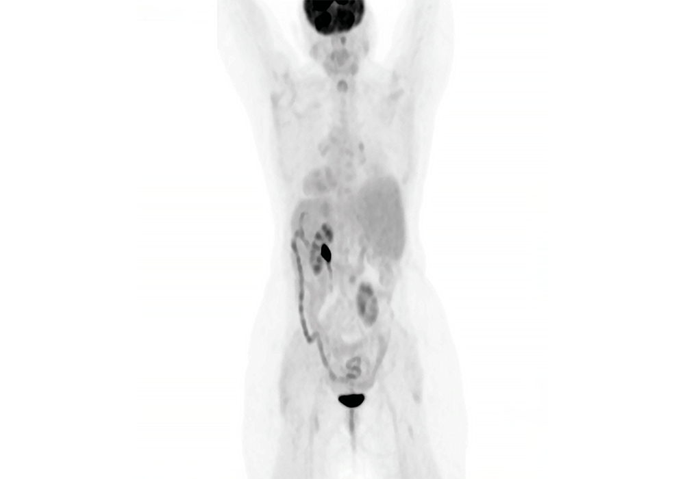
    After 2 months on a selective RET inhibitor, the patient had her first restaging PET-CT, which revealed an almost complete resolution of her mediastinal lymphadenopathy and improvement of her previous right-sided consolidation. Subsequent scans demonstrated deepening of this response without evidence of PET avidity.
    The patient has tolerated therapy well with only intermittent nausea, which has not required antiemetic therapy, dose holding, or even dose reduction. She continues on therapy almost 2 years later with continued response on radiographic imaging.
    So, in summary, we have a patient with a RET fusion that was treated eventually with a selective RET TKI and has done quite well. And I think there are several key takeaways from this case. The first is that all of our patients in the metastatic setting and now even in earlier stages should be getting comprehensive genomic testing. And this is critical to identify these patients who can benefit from targeted therapy, such as in this case, and also select patients that will not benefit from immune checkpoint inhibitors. The advantage of this is that we're getting patients who potentially can have higher responses and durable responses. If you have a patient where you can't wait for the testing to come back, that's fine. But what I would recommend in those situations is to hold the immunotherapy for the first cycle and wait for that molecular testing to come back if you're worried about disease burden. There's a couple reasons for doing this. One is that if we give targeted therapy after immunotherapy, there can be a high side effect profile. And two, you may cause unnecessary autoimmune side effects like we saw in this patient
    I want to thank everybody for listening to this patient case presentation. We hope the information provided here will have a positive impact on your treatment approach for patients with advanced or metastatic non–small cell lung cancer.

    Move image slider tool to see the after treatment scan

    PET scan of patient after treatment
    PET scan of patient before treatment
    • Judicious use of IHC to conserve tumor tissue for molecular studies, especially in patients with advanced disease
    • Broad molecular profiling using a validated test to minimize tissue use and potential wastage and assess all recommended biomarkers in all appropriate patients with NSCLC
    • Patients with these oncogenic mutations should receive treatment with targeted agents


    Welcome. My name is Luis Raez. I am the chief scientific officer and medical director of Memorial Cancer Institute at the Memorial Healthcare System. We are the third largest public healthcare system in the United States. We are situated in South Florida. Also, I'm still the Director of the Thoracic Oncology program. I am a lung cancer doctor by career and my focus of research is based on the molecular targets and liquid biopsies.
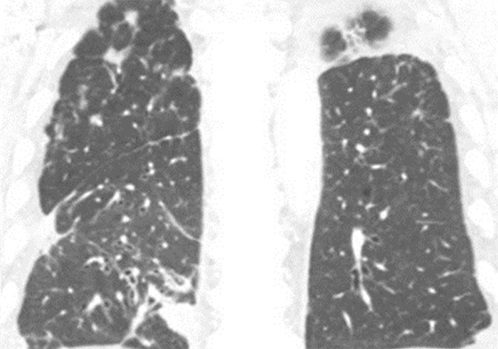
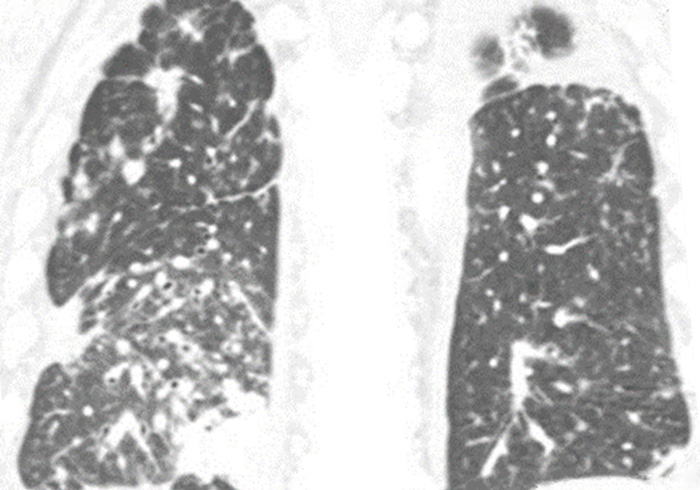
    Today, we’ll be discussing the case of a patient who was referred to my care. The patient was an 80-year-old male with stage IV non–small cell lung cancer. At diagnosis, his adenocarcinoma was infiltrating both lungs, as you can see in the pictures, and had metastasized to his bones. Also, after 4 years of treatment after diagnosis, the patient was found to have developed brain metastases.
    Now we’ll review the patient’s story that brought him to this point as it was given to me when he transferred to my care.
    The patient reported to never have smoked, used smokeless tobacco products, or partaken in alcohol or drug use.
    You can see in the table that the patient has some laboratory studies that we're displaying. The patient had weakness and malaise only as symptoms. So, the patient was still in good performance status and the patient was not very symptomatic as you can see. He was a senior citizen certainly, but he was very motivated looking for opportunities of therapy because as we can see later, he has failed several standard therapies.
    The best molecular assessment probably can be done only with next-generation sequencing, because we know in metastatic non–small cell lung cancer, we have now 10 actionable genetic aberrations that need to be evaluated. So, there is no way nowadays that other technologies, like PCR, FISH, immunohistochemistry, can give us the answer because it's a matter of cost. It's a matter of time. That is why the only comprehensive or complete genetic analysis comes to doing next-generation sequencing in tissue or in blood or in both.
    After his initial diagnosis of stage IVb non–small cell lung cancer, liquid and tissue samples were sent for molecular biomarker testing. The liquid biopsy results came back as negative for any actionable mutations or genetic aberrations. However, these results would not be confirmed by the
    tissue-based NGS test since the sample was deemed as “quantity not sufficient.” Immunohistochemistry of the tissue sample revealed low PD-L1 levels. Maybe some of us will have tried to pursue a little bit further and trying to get an answer from tissue because, as we said before, none of these technologies either liquid or tissue are 100% accurate yet, and that is why we need them to complement each other.

    Because the patient was ruled to be ineligible for targeted therapies and immunotherapy was not part of the standard of care yet, he was started on palliative chemotherapy with Carboplatinum and Paclitaxel, plus Denosumab for his bone metastases.
    The best course of action at progression in this patient, today, probably will be to do another genetic analysis of the tumor, either by a liquid biopsy or a tissue biopsy, if possible. This patient was treated some years ago. That is what we will see in the next slides, the steps that he went for his therapy.
    After the patient had disease progression with the frontline therapy, Nivolumab was added as a second-line therapy, at that time was the standard of care, until the patient had disease progression again.
    Over the next year-and-a-half to 2 years, the patient was cycled through 4 additional chemoimmunotherapy regimens as his disease continued to progress. It was on his last line of therapy, Gemcitabine, that brain metastases were found, and he was treated with whole brain therapy.
    It was at this point that the patient was transferred into my care and his lung tumor was sent for tissue-based NGS testing, and the result came back positive for the most popular RET fusion, the KIF5B, which is an actionable biomarker in non–small cell lung cancer.
    And we had a clinical trial open for a RET inhibitor in our cancer center, because this was two years before any RET inhibitor approval, and we were able to enroll the patient in the clinical trial.
    The patient came to us because he was running out of options. He already has tried 6 different lines of therapy including 5 chemotherapeutic agents and immunotherapy. So basically, for him the standard of care probably had only 1 or 2 more chemotherapeutic drugs available with a very low chance of success at that time in the standard of care.
    At this point, the patient was started with a RET inhibitor at a conventional dose.
    After 3 months of treatment, the patient’s CT scans showed marked improvement of the infiltrates in the right and left lungs.
    On the selective RET inhibitor, the patient experienced 18 months of progression-free survival with a continued quality of life that was unaltered by side effects. Additionally, there were no major alterations to his laboratory work that could not have been controlled by adjusting the dose of his pre-existing medications.
    You are talking about a patient that has metastatic lung cancer who has failed 6 lines of palliative therapy before. So, 18 months of progression-free survival is a lot for him.
    You know, there are several key takeaways here. First of all, the judicious use of immunohistochemistry to conserve more tissue for molecular studies, especially in patients with advanced disease. If this is the second, third or fourth biopsy that you're doing in your patient, please notify the pathologist that we don't have to be using a lot of material to prove that this is lung cancer.
    Another important takeaway is to test it for all actionable biomarkers. I remember we said this at the beginning of the talk. We need to do a comprehensive next-generation sequencing in the tissue or in the blood.
    But this also has to be done, as I said, in the second, third, or fourth time that the patient fails treatment so we can identify new genetic markers that can be used to rescue the patient. And if we do that, patients will be having targeted agents as treatment.
    Thank you for listening to this patient case presentation. We hope that the information provided here will have a positive impact in your treatment approach for patients with advanced or metastatic non–small cell lung cancer.

    Move image slider tool to see the after treatment scan

    CT scan of patient after treatment
    CT scan of patient before treatment
    • Perform broad molecular profiling using a validated test to minimize tissue use and potential wastage and assess a for actionable genetic biomarkers
    • Have molecular testing results for the actionable oncogenic mutations before starting systemic therapy combined with immunotherapy
    • Treat patients with oncogenic driver mutations with targeted first-line therapy for that oncogene


    Welcome. My name is Puja Arora. I am a community hematologist-oncologist with University Hospitals Seidman Cancer Center located west of Cleveland at
    St. John Medical Center. I see most tumor types, and my practice is largely made up of breast, lung and GI malignancies, but I also see malignant and benign hematology.

    Today, we’ll be discussing the case of a patient who I initially met as a second opinion. The patient is a female, 53 years of age with stage IV non–small cell lung cancer.
    When I met this patient, she had already consented to chemoimmunotherapy, but she was seeking a second opinion before beginning treatment. Now, we’ll review the patient’s history leading up to this point after she was transferred to my care.
    The patient was previously healthy with no significant past medical history. She was a never smoker and reported no alcohol use. She did report asbestos exposure through her work with a nonprofit organization though could not provide details.
    She initially presented to the emergency room with shortness of breath to the point she could not walk to her mailbox. A CT of the chest on arrival revealed a soft tissue density in the right side of the mediastinum at the mid-right hemithorax and mediastinal lymph nodes along with a T12 lesion and bilateral pleural effusions.
    Pathology from thoracentesis revealed adenocarcinoma of pulmonary origin.
    After the patient’s initial diagnosis of stage IV non–small cell lung cancer, tissue samples were sent for molecular biomarker testing. A lung cancer hotspot gene panel came back negative for variants in BRAF, EGFR, HER2, KRAS, and MET. Immunohistochemistry of the tissue sample revealed low levels of PD-L1 expression with a tumor proportion score of 11%-20%.
    The biomarker testing was completed at a well-known outside institution using their in-house assay, which only evaluated
    5 of the 9 actionable genomic markers in non–small cell lung cancer.

    While these 5 markers were negative, it was not comprehensive genomic profiling and, in this case, led to selection of a therapeutic regimen without having all of the necessary information to make that decision.
    The patient’s original physician recommended chemoimmunotherapy with carboplatin, pemetrexed and pembrolizumab. Although the patient consented to this treatment plan, she sought a second opinion at my clinic before initiating therapy.
    Given that the patient was a nonsmoker and did not have the typical risk factors for lung cancer, it was even more important to have comprehensive genomic profiling prior to initiating any therapy.
    After reviewing her case and knowing there may be some delays in getting tissue from an outside institution, I elected to obtain a liquid biopsy the day I saw her to allow for a quick turnaround time. Results came back with a RET fusion, which is an actionable biomarker in non–small cell lung cancer.
    The patient was now eligible for a targeted therapy, and after some discussion, she decided to move forward with a selective RET inhibitor. She initiated treatment within
    1 month of her original diagnosis.

    After 3 months of treatment with a selective RET inhibitor, the patient’s PET scan showed an almost complete response, with complete resolution of the
    T12 lesion and pleural effusion.

    Furthermore, the patient reported alleviation of her disease symptoms within 2 weeks of treatment initiation.
    She no longer had any shortness of breath and was able to stop taking all of her pain medications.

    The patient continues to experience progression-free survival with a quality of life that was unaltered by side effects.
    Here are some key takeaways from this case. This case highlights why it is so important to have comprehensive genomic profiling before initiating any therapy. Point 3 demonstrates the number of genetic variants that are now available and should be looked for in non–small cell lung cancer. This patient could have missed out on an opportunity to have a targeted therapy. Her quality of life was preserved, and she continues to live life working full time and travelling with her spouse without having any limitations from her cancer.
    Thank you for listening to this patient case presentation. We hope the information provided here will have a positive impact on your treatment approach for patients with advanced or metastatic non–small cell lung cancer.

    Move image slider tool to see the after treatment scan

    PET scan of patient after treatment (front view)
    PET scan of patient before treatment (side view)
    Treatment Roadmap for HCPs

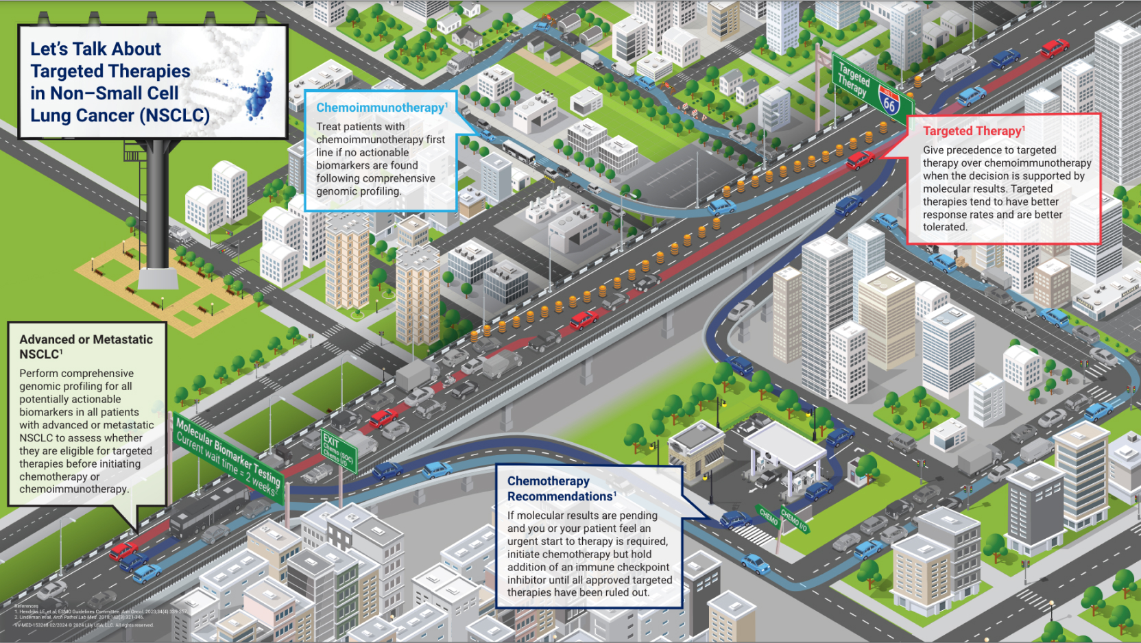
    The roadmap is designed to be used in discussions HCPs and their patients with NSCLC to facilitate informative and meaningful conversations that will help prepare patients to begin their treatment journeys. Discussion topics include guideline-concordant molecular biomarker testing, as well as recommendations on traditional chemotherapy, chemoimmunotherapy, and oncogene-targeted therapy treatment options for these patients. See the “Related Resources” section to download the roadmap, discussion guide, and accompanying video for your patients.


    Every patient diagnosed with non-small cell lung cancer is embarking on a personal treatment journey.

    The decisions they make can impact not only their outcomes, but the kinds of obstacles they will face along the way.

    While providing medical explanations and trusted advice, you are often your patient’s main source of information. The tone you set and guidance you provide impacts patient decision making.

    One way to help patients make the best decision for themselves is to provide as much insight and data as possible. Molecular biomarker testing can provide the most appropriate roadmap to navigate treatment options, but the wait time for results can be difficult.

    One way to help patients make the best decision for themselves is to provide as much insight and data as possible. Molecular biomarker testing can provide the most appropriate roadmap to navigate treatment options, but the wait time for results can be difficult.

    Do they stay on this route and wait for traffic to clear or do they take an exit and search for a path around the backup?

    For a patient, waiting to receive molecular biomarker test results may seem as frustrating as the wait in traffic. But with much higher stakes. When multiple treatment paths can be taken, each requires careful planning for optimal outcomes.

    Let’s look at three possible scenarios.

    When the dark blue car encounters the traffic jam, they’re anxious and feel the need to begin treatment immediately, instead of waiting for biomarker results.

    For the driver, feeling like they need to refuel may lead them to exit and fill up at a nearby gas station before returning to the highway.

    Similarly, starting a patient on a round of chemotherapy while waiting for biomarker results may achieve a compromise on treatment path with minimum impact on the efficacy of potential targeted therapies later.

    Here, the light blue car also exits the highway to search for an alternate route. To keep moving, they choose a winding path through the city before eventually re-entering the highway later.

    However, city streets come with their own obstacles, which can be as frustrating as waiting in the traffic jam and are not necessarily better routes to the driver’s destination.

    Similarly, patients may want to explore treatment paths they can begin immediately instead of waiting for biomarker results. For patients eligible for immunotherapy, that treatment route may initially look better because it can be started quickly, but it may make them ineligible for later targeted therapy.

    The red car, however, stays on the highway and waits through the traffic jam. It may seem tedious, but sometimes waiting for traffic to clear is the fastest way through.

    Although molecular biomarker testing may come with a wait, results provide the most complete roadmap for navigating treatment.

    Patients who are positive for an actionable biomarker can be given a targeted therapy, which tend to be better tolerated and have greater efficacy, for a clear path ahead as they navigate their cancer journey.

    There are no “right” answers. But your guidance can help patients feel supported as they make difficult decisions.

    You can find This Treatment Roadmap and other patient resources to help facilitate discussions with patients about treatment plans by scanning the QR codes.

    Download the Let's Talk About Targeted Therapies in Non–Small Cell Lung Cancer Roadmap.

    Preview of the downloadable roadmap.


    Managing Disease Progression

    Both intrinsic and acquired resistance remains a challenge when patients are treated with TKIs, with most patients experiencing disease progression. Intrinsic mechanisms are present before therapy, whereas acquired mechanisms are induced after therapy is initiated. Common mechanisms of resistance to TKIs include101-104:

    • TKI domain or other drug-binding site mutations
    • Downstream signaling effector mutations
    • Bypass signaling pathways

    For patients with a documented prior actionable alteration in whom the disease has progressed with therapy, retesting should be done exclusively on rebiopsy specimens of a progressing lesion. If a biopsy is performed on a suspected bony metastasis, it is critical that decalcification be avoided because some methods of decalcification can preclude any subsequent molecular testing.81


    Post-diagnostic Use of Biopsies105

    Post-diagnostic Use of Biopsies
    chevron-filled-down View image description

    Related Resources

    Downloadable PDFs

    Download PDF Medical Answer PDF Document Created with Sketch. Infographic: Minimizing Turnaround Time for Molecular Testing Among Patients With NSCLC

    This infographic is intended to educate healthcare providers on barriers and solutions for minimizing turnaround time for molecular testing.

    Download PDF Medical Answer PDF Document Created with Sketch. Let's Talk About Targeted Therapies in Non–Small Cell Lung Cancer Roadmap

    This roadmap is an educational tool about treatment pathways for patients diagnosed with advanced or metastatic non–small cell lung cancer (NSCLC), designed to be used in discussions between healthcare professionals (HCPs) and their patients.

    Download PDF Medical Answer PDF Document Created with Sketch. HCP Roadmap Discussion Guide

    This discussion guide is designed to be used with the Let's Talk About Targeted Therapies in NSCLC roadmap to facilitate conversations between HCPs and their patients with NSCLC.

    Download PDF Medical Answer PDF Document Created with Sketch. Patient Roadmap Conversation Guide

    This discussion guide is designed to be used with the Let's Talk About Targeted Therapies in NSCLC roadmap to facilitate conversations between patients with NSCLC and their HCPs.


    Question 01 – What prompted you to consider internalizing CGP?

    So comprehensive genomic profiling is a technique that really tells us a lot about a tumor in one single test. It combines information that was previously only available by combining several different single gene tests.

    The reason why we want a test like comprehensive genomic profiling is it represents the highest standard of care that we can provide to our patients. So for us as pathologists this is the best thing we can do from inside the laboratory to help a patient on their cancer journey. It’s also the best level of support that we can provide to our clinicians like our medical oncologists or our surgeons who are taking care of these patients.

    Prior to the widespread availability of techniques that provide comprehensive genomic profiling, we were really using single gene tests. And these were great when that's all we had, And the issue, though, and the problem with them is that they provide a very limited amount of information.

    I look at these as if they are yes/no questions. So, for instance, in a disease like lung cancer, we might want to ask, “Does this patient have an EGFR mutation?” If the answer is yes, then they can go on to a targeted therapy towards that particular mutation.

    But if the answer is no, then we have to ask another question. We have to ask, “Does this patient have an ALK fusion?” or “Does this patient have a ROS fusion?”

    And each time if the answer is no, we’re no further ahead than we were before. And each time we ask a question we lose a little bit of tissue. Eventually the tissue will run out, and we’re not able to ask any further questions.

    With comprehensive genomic profiling, it’s almost that right out of the gate we can ask a different question that’s open ended. We can say, “What is driving this tumor?” When we have an answer to that question in hand, we’re able to provide the most informed therapeutic decisions right out of the gate.

    Question 02: What are the advantages of rapid NGS, and how has it changed workflows at your hospital system?

    So when it comes time to delivering biomarkers, in particular next generation sequencing–based biomarkers for cancer care, there is an immense need for speed. And this is perceived from patients, from oncologists,

    and in order for us to understand that from within the laboratory, we really need to understand what’s going on with our patients and a little bit about these diseases. Lung cancer, I think, provides the best example of why these results need to happen very fast.

    In Canada, about 50% of lung cancer patients will present at an advanced stage, a stage IV, requiring a systemic therapy. These patients are very sick, and they tend to present in extremis, requiring treatment right away.

    In fact, we know that the death rate of advanced non–small cell lung carcinoma prior to treatment is about 4% per week. Also, in a publicly funded healthcare system like in Canada, it costs us about $400 a week or more to provide supportive care to these patients in the period in between their diagnosis and when they first receive therapy.

    So it is paramount that we get these results quickly, as without a complete set of biomarker results, it is impossible for an oncologist to decide on the most appropriate treatment for a lung cancer patient.

    I practice in a community hospital, which is where the majority of cancer patients are diagnosed and treated in Canada and in many similar jurisdictions around the world. Traditionally, we send our biomarkers out to another academic hospital and using that route, it takes 6 to 8 weeks to receive results.

    When we insourced biomarker testing using a rapid method, the turnaround time went from 64 days on average to 3 days on average. The difference was immense.

    This meant that the majority of cancer patients were going to have their results available at the time when they first met with their medical oncologists. So that means when the have that first encounter, the oncologists could make a critical decision as to if this patient was going to receive a targeted therapy and what that appropriate targeted therapy would be or if they were going to receive chemotherapy or immunotherapy.

    And without that information present at that first oncology visit, the visit becomes a waste. It becomes impossible to start the patient on any form of treatment.

    Question 03: How has your institution used rapid NGS to improve access for patients? How has the use of rapid NGS changed patient care?

    So for modern-day cancer treatment, precision therapy is really where it’s at, and this is what everybody is looking for in terms of the cancer treatment that they want. Next-generation sequencing is the cornerstone of precision medicine, and having that next-generation sequencing available is a necessary precursor to get our patients eligible to receive that precision therapy.

    When NGS service becomes a part of our institution and is offered within our four walls, it enables us to make sure that 100% of our patients are going to have access to that and are going to have their tumors on that next-generation sequencer in order to set them up for precision therapy down the line.

    When we have this ability in our hospital, we can really control the workflow in a way that it’s patient centric.

    We’re no longer sending that material out in the mail. We’re no longer dependent on outside institutions faxing or emailing those results back, which can be unreliable.

    And instead, we’re providing that result direct to our oncologists.

    When the technique is available and accessible to our oncologists, they can order it whenever they want, wherever they want.

    But also, a lot of other benefits come out of the woodworks. Our pathologists who are making that diagnosis all of a sudden can order the same test, too, and that actually helps refine what they’re seeing under the microscope and make sure that we’re starting off with more accurate diagnoses right out of the gate.

    Question 04: Is rapid NGS cost-effective?

    Yes. Rapid NGS is certainly cost-effective, but it can be a little hard to appreciate that. When we think about the costs of NGS, we often get stuck on the costs of a next-generation gene sequencer, and these can range anywhere from $50,000 to over a million dollars.

    And then we can often focus on the cost of the consumables, like the little plastics and chemicals that make these machines go. But in my experience, the human resources required to run these instruments is actually one of the biggest costs that we face. So these are the pathologists in your lab, but more importantly, the technologists who actually run this technology and produce the results.

    When you use a rapid NGS, these techniques are largely automated. There’s a lot of robotics that is replacing steps that used to be done manually. And so when we move to a rapid technique, we’re not only making these results come out faster, but we’re really paring down the amount of human resources that are required in order to produce that NGS result. So there’s a lot cost savings there for the laboratory.

    But another thing we need to do is take a step back and not look at costs just in the laboratory. Cancer is a health systems issue. So introducing this within the lab can produce a lot of cost savings in the oncology clinic but also in the pharmacies.

    We know that getting on the correct treatment right away is essential, and if we have to refine that by switching therapies after biomarker results show up late, there’s a huge cost associated with that. In addition, simply having those oncologist-patient interactions being productive and not requiring second appointments or return visits produces another huge cost saving for the healthcare system as a whole.

    Question 05: What would you say to institutions that do not have in-house CGP?

    I think that the way we view this has really changed over time. Maybe 10 years ago, this might have been a new entity or something that’s emerging out of the research world that is going to one day have a role in clinical care. But right now, next-generation sequencing and comprehensive genomic profiling–these are standards of care. And so if this is not available, it means that your patients are not likely getting the most appropriate treatment.

    So when you bring NGS to your community or to your hospital, it’s a really major undertaking. For a pathologist, this is something that can define your career. But it’s important to understand that it’s not just a laboratory tool.

    Pathologists should not be left on their own to do this job. We really need help from our oncologists, surgeons, and even our patients and patient advocacy groups to all come to our hospitals together to make this happen.

    But when it’s all done, at the end of the day, the benefits are immense, and they’re not just for the laboratory. The entire hospital feels the benefit of this.

    It improves the way clinical oncology works in the clinics; it improves the way surgical practices work. It improves the diagnostic yield of biopsies. And the benefits even go beyond the hospital. They're felt by the entire community,

    and it’s very satisfying to know that it wasn’t just the laboratory, but the entire team helped bring in this new tool and that this new tool is helping us achieve that highest level of cancer care.

    Question 06: How do you see the widespread implementation of rapid NGS improving patient care in the future?

    So when we talk about rapid NGS, it’s certainly fast, and the benefits come from the speed. But how we achieve that rapid nature of the NGS is by a large degree of automation. And when we automate the process of NGS, this makes it easier to perform in certain areas that were not typically conducive to NGS testing in the past.

    So when we take a very complicated process and put it under the control of a robot, we can perform this task in community hospitals, in rural regions, and other areas that don’t have the traditional expertise required to run a complicated NGS service.

    So by automating our NGS, we can achieve what we call a point-of-care–style testing and make this very simple and easy to use in regions where that NGS information is going to be most helpful to cancer patients.

    So in the future I see point-of-care NGS, or automated or rapid NGS, playing a major role in those underserved regions in community hospitals in remote and rural locations, bringing that idea of precision cancer care to life in regions where it was not previously possible.

    Insourcing Rapid NGS in a Community Hospital with Dr. Brandon Sheffield

    Dr. Brandon Sheffield discusses insourcing rapid NGS in a community hospital and its role in precision medicine.


    INTRODUCTION

    Dr. Kolesar: Hi, I’m Jill Kolesar. I'm a clinical pharmacologist and a professor at the University of Kentucky College of Pharmacy. I'm also the director of our molecular tumor board and I came to the University of Kentucky, in 2016, to start a precision medicine program and the molecular tumor board is a big part of that. One of our goals at the Markey Cancer Center is to provide the molecular tumor board to all of our academic medical oncologists as well as a community medical oncologists across our state.

    Dr. Thind: Hi, I’m Ravneet Thind. I'm a medical oncologist and I work at a community hospital in eastern Kentucky. I have known Jill for six, seven years, and for the last year, or so, we have worked very closely in an attempt to increase awareness and sort of emphasize the importance of next generation sequencing and molecular tumor boards in our community.

    QUESTION 01: What was the impetus for setting up the MTB at UK Markey Cancer Center?

    Dr. Kolesar: The molecular tumor board was established with the support of the Markey Cancer Center administration due to the recognition of very low molecular testing rates in our state. We started at the Markey Cancer Center and recently expanded into our community medical oncology practices.

    As the only NCI-designated comprehensive center in the state, we are partnering with our local community medical oncology practices to advance the use of molecular testing and targeted therapy.

    Dr. Thind: Comprehensive genomic profiling has become an integral part of our practice to better understand the biology of tumors. Community oncologists are increasingly faced with advanced molecular diagnostic data yet have minimal training in genomics.

    This creates a void between clinical practice and technologic potential, and I think a molecular tumor board is a great way to bridge this gap.

    QUESTION 02: What are the advantages of an MTB?

    Dr. Kolesar: We implemented a molecular tumor board with the intention of increasing the rates of molecular testing, which we anticipated would decrease turnaround time for testing, increase access to clinical trials, and ultimately, more patients receiving biomarker-informed treatment decisions.

    Dr. Thind: Due to the general cytotoxicity of conventional chemotherapy, focus has now shifted to novel therapeutic targets that can be exploited based on genomic data and molecular tumor board recommendations. So as Jill mentioned previously, molecular tumor boards can increase guideline-concordant testing and decrease turnaround time for molecular testing, which enables biomarker-informed decision-making and optimizes therapeutic options for our patients.

    Dr. Kolesar: Dr. Thind, as you can see in this figure from our case-control study, we showed that patients with non–small cell lung cancer who had their cases reviewed by a molecular tumor board had improved overall survival compared to those who did not, regardless of whether they were treated in an academic setting or a community.

    Question 03: What elements were necessary for extension of the MTB to rural community sites?

    Dr. Kolesar: I think the most important part was really to make it easy for sites to participate. So to do that, we held a virtual meeting, and we had a molecular tumor board coordinator that managed the cases referred from the community sites.

    Our coordinator interacts with clinic staff from the treating physician office to gather up the notes and NGS reports for review and to provide the written recommendations back. They also coordinate the virtual meeting.

    Dr. Thind: Yes, I agree. I think having a molecular tumor board–dedicated navigator has been a game changer. Along with excellent communication and support from the University of Kentucky, with clear and precise explanation of the entire process.

    Dr. Kolesar: I think it was also really helpful to demonstrate the clinical benefit showing that patients with molecular tumor board review had better outcomes.

    And having wide clinical and genomic expertise, with evidence grading were also critical to the success of the molecular tumor board.

    Question 04: What would you say to institutions that don’t currently have an MTB but are weighing the investment of resources to create a dedicated program?

    Dr. Thind: Tumor boards are a long-standing tradition in oncology, and I think molecular tumor board is a step above this.

    As molecular diagnostic testing is becoming more and more sophisticated and complex, molecular tumor boards can bring relevant expertise to this rapidly emerging field and are crucial for patient care.

    In my opinion, molecular tumor board is the road that leads to precision medicine in oncology. It is quite simply the best thing for your patients.

    Dr. Kolesar: I think a molecular tumor board is a win-win for everyone. It brings medical oncologists, pathologists, clinical pharmacists, and genetic counselors together, providing the opportunity for true clinical collaboration.

    But most importantly, patients live longer, and when patients live longer, providers and health systems have more time to care for patients and achieve better outcomes.

    For institutions without a current molecular tumor board and without additional resources to start one, I would encourage them to partner with and learn from existing molecular tumor boards. Presenting cases is a great initial step; existing molecular tumor boards would appreciate the clinical input and can help support other sites with infrastructure.

    Question 05: How do you see the widespread implementation of MTBs improving patient care in the future?

    Dr. Kolesar: Right now, molecular tumor boards are a labor of love and require dedication and commitment from a large interprofessional team.

    Our group and other groups are tracking patient-level outcomes with MTBs and developing artificial-intelligence applications to support the widespread implementation and use of molecular tumor boards.

    Our AI model is trained on patient characteristics and outcomes on molecular tumor board–recommended therapy, and we plan to use it to predict the best therapies for our future patients. I think widespread application of molecular tumor boards, supported by AI, has the potential to transform and democratize cancer care and dissemination of precision medicine.

    Expanding Molecular Tumor Boards to Community Settings with Drs Jill Kolesar and Ravneet Thind

    Drs Jill Kolesar and Ravneet Thind discuss expanding molecular tumor boards to community settings and the clinical benefits that follow.


    Minimizing turnaround time for molecular testing among patients with non-small cell lung cancer is key to increasing access to targeted therapies for patients. Less than 40% of eligible patients receive an appropriate targeted therapy, with 31% of this patient loss due to barriers with biomarker test ordering, communication of findings, and subsequent treatment decisions.

    Examining common barriers and solutions may help standardize molecular biomarker testing and improve patient access to targeted therapies. To this end, we have identified turnaround time-related challenges across the testing process, presented here as pre-analytic, analytic, and post-analytic barriers. For each barrier, multiple implementable solutions and key takeaway points are provided to help clinicians and pathologists work together more efficiently and effectively. To learn how these solutions can help minimize turnaround time for molecular biomarker testing for your patients with non-small cell lung cancer, download this infographic from education.lillymedical.com.

    Minimizing Turnaround Time for Molecular Testing Among Patients With NSCLC

    This video summarizes the infographic on barriers and solutions for minimizing turnaround time for molecular testing.


    Every patient diagnosed with non-small cell lung cancer is embarking on a personal treatment journey.

    The decisions they make can impact not only their outcomes, but the kinds of obstacles they will face along the way.

    While providing medical explanations and trusted advice, you are often your patient’s main source of information. The tone you set and guidance you provide impacts patient decision making.

    One way to help patients make the best decision for themselves is to provide as much insight and data as possible. Molecular biomarker testing can provide the most appropriate roadmap to navigate treatment options, but the wait time for results can be difficult.

    One way to help patients make the best decision for themselves is to provide as much insight and data as possible. Molecular biomarker testing can provide the most appropriate roadmap to navigate treatment options, but the wait time for results can be difficult.

    Do they stay on this route and wait for traffic to clear or do they take an exit and search for a path around the backup?

    For a patient, waiting to receive molecular biomarker test results may seem as frustrating as the wait in traffic. But with much higher stakes. When multiple treatment paths can be taken, each requires careful planning for optimal outcomes.

    Let’s look at three possible scenarios.

    When the dark blue car encounters the traffic jam, they’re anxious and feel the need to begin treatment immediately, instead of waiting for biomarker results.

    For the driver, feeling like they need to refuel may lead them to exit and fill up at a nearby gas station before returning to the highway.

    Similarly, starting a patient on a round of chemotherapy while waiting for biomarker results may achieve a compromise on treatment path with minimum impact on the efficacy of potential targeted therapies later.

    Here, the light blue car also exits the highway to search for an alternate route. To keep moving, they choose a winding path through the city before eventually re-entering the highway later.

    However, city streets come with their own obstacles, which can be as frustrating as waiting in the traffic jam and are not necessarily better routes to the driver’s destination.

    Similarly, patients may want to explore treatment paths they can begin immediately instead of waiting for biomarker results. For patients eligible for immunotherapy, that treatment route may initially look better because it can be started quickly, but it may make them ineligible for later targeted therapy.

    The red car, however, stays on the highway and waits through the traffic jam. It may seem tedious, but sometimes waiting for traffic to clear is the fastest way through.

    Although molecular biomarker testing may come with a wait, results provide the most complete roadmap for navigating treatment.

    Patients who are positive for an actionable biomarker can be given a targeted therapy, which tend to be better tolerated and have greater efficacy, for a clear path ahead as they navigate their cancer journey.

    There are no “right” answers. But your guidance can help patients feel supported as they make difficult decisions.

    You can find This Treatment Roadmap and other patient resources to help facilitate discussions with patients about treatment plans by scanning the QR codes.

    PMDx NSCLC Roadmap HCP Overview Video

    This video is designed to be used with the Let's Talk About Targeted Therapies in NSCLC roadmap to facilitate conversations between HCPs and their patients with NSCLC


    Dan, a delivery driver, was recently diagnosed with non-small cell lung cancer and his mind is a snarl of piled up thoughts. One of his biggest concerns is treatment options.

    His doctor wants to do a molecular biomarker test to see if Dan is eligible for any targeted therapies. He’s agreed, but it could be weeks before they have the results, and Dan is nervous about waiting to start treatment.

    During the conversation about treatment planning, Dan’s doctor shared a treatment roadmap that breaks down three possible scenarios.
    We’ve all been in a highway traffic jam and had to make a decision. Should we stay on the highway and wait for traffic to clear, or do we exit and search for a path around the backup?


    For a patient like Dan, waiting for molecular biomarker test results may seem as frustrating as the crawl through traffic. But with much higher stakes. When multiple treatment paths can be taken, each requires careful thought and planning.

    Consider three possible scenarios from the Treatment Roadmap.

    When the dark blue car encounters the traffic jam, waiting makes them anxious and they feel the need to take action. So, they exit to refuel before getting back onto the highway.

    Similarly, patients may sometimes want to explore treatment paths that they can begin immediately instead of waiting for biomarker results.

    For some, starting a round of chemotherapy while waiting for results may achieve a compromise that still allows for possible targeted therapies later.

    The light blue car also exits the highway to search for an alternate route. To keep moving, they choose a winding path through the city before eventually re-entering the highway.

    However, city streets come with their own obstacles, which can be as frustrating as waiting in traffic and are not necessarily better routes to the driver’s destination.

    In this case, patients may want to explore more involved treatment paths they can begin immediately instead of waiting for biomarker results. For patients eligible for immunotherapy, that treatment route may look better at first because it can be started quickly.

    But it may make them ineligible for targeted therapy later.

    The red car stays on the highway and waits through the traffic jam. It may be frustrating, but sometimes waiting for traffic to clear may be the fastest way through.

    Although molecular biomarker testing might come with a wait, results provide the most complete roadmap for navigating treatment.

    Patients who are positive for an actionable biomarker can be given a targeted therapy, which tend to be better tolerated and have greater efficacy, for a clear path ahead as they navigate their cancer journey.

    As a driver, Dan knows destinations often have more than one possible route, but, depending on the situation, they’re not all optimal. His treatment is going to be the same way. But at least he has somewhere to start.

    Talk to your care team about how the Treatment Roadmap can help you consider the treatment routes you've discussed.

    PMDx NSCLC Roadmap Patient Overview Video

    This video is designed to be used with the Let's Talk About Targeted Therapies in NSCLC roadmap to facilitate conversations between patients with NSCLC and their HCPs

    ADC=antibody drug conjugate; AKT=protein kinase B; ALK=anaplastic lymphoma kinase; ARAF=A-rapidly accelerated fibrosarcoma; ATP=adenosine triphosphate; ATR=ataxia telangiectasia and Rad3-related protein; AXL=tyrosine-protein kinase receptor UFO; BAD=B-cell lymphoma-extra large/-2 associated death promoter; BCL-X=B-cell lymphoma-extra large; BRAF=B-rapidly accelerated fibrosarcoma; ChIP=chromatin immunoprecipitation; CMS=Centers for Medicare & Medicaid Services; CNV=copy number variation; CR=conserved region; CRAF=C-rapidly accelerated fibrosarcoma; ctDNA=circulating tumor deoxyribonucleic acid; DAG=diacylglycerol; dMMR=MMR deficient; EBUS=endobronchial ultrasound bronchoscopy; EGFR=epidermal growth factor receptor; EML4=echinoderm microtubule-associated protein-like 4; EMR= electronic medical records; ERBB2=erythroblastic oncogene B 2; ERK=extracellular signal-related kinase; FISH=fluorescence in situ hybridization; FNA=fine needle aspiration; FN3=fibronectin type III; GAB=Grb2-associated binding scaffold protein; GDNF=glial cell line-derived neurotrophic factor; GDP=guanosine diphosphate; GF=growth factor; GFRα=GDNF family receptor-α; GI=gastrointestinal; GTP=guanosine triphosphate; HCP=healthcare professional; HER2=human epidermal growth factor receptor 2; HGF=hepatocyte growth factor; ICI=immune checkpoint inhibitor; IHC=immunohistochemistry; IPT=immunoglobulins, plexins, and transcription factors; IP3=inositol trisphosphate; JAK=Janus kinase; JNK=JUN N-terminal kinase; KIF5B=kinesin family member 5b; KRAS=Kirsten rat sarcoma; LDL=low-density lipoprotein; LRR=leucine-rich repeat; MAM=meprin/A5/Mu; MAPK=mitogen-activated protein kinase; MEK=mitogen-activated protein kinase; MET=mesenchymal-epithelial transition; MMR=mismatch repair; MSI=microsatellite instability; mTOR=mammalian target of rapamycin; NCOA4=nuclear receptor coactivator 4; NF-κB=nuclear factor kappa B; NGS=next-generation sequencing; NSCLC=non–small cell lung cancer; NT=neurotrophic; NTRK=neurotrophic tyrosine receptor kinase; PAK=P21-activated kinase; PCR=polymerase chain reaction; PDK1=pyruvate dehydrogenase lipoamide kinase isozyme 1; PD-L1=programmed death-ligand 1; PIP2=phosphatidylinositol biphosphate; PI3K=phosphatidylinositol 3-kinase; PKA=protein kinase A; PKC=protein kinase C; PLC=phospholipase C; PSI=plexins, semaphorins, and integrins; QNS = quantity not sufficient; RAC1=Ras-related c3 botulinum toxin substrate 1; RAF=rapidly accelerated fibrosarcoma; RAS=rat sarcoma; RET=rearranged during transfection; RHO=rhodopsin; ROSE=Rapid OnSite Evaluation; ROS1=ROS proto-oncogene 1; SGT= single gene test; SNV=single-nucleotide variant; SOS=Son of Sevenless; STAT=signal transducer and activator of transcription; TAT=turnaround time; TFG=tropomyosin-receptor kinase-fused gene; TIAM1=T-lymphoma invasion and metastasis-inducing protein 1; TKI = targeted kinase inhibitor; TM=transmembrane; TMB=tumor mutation burden; TRIM=ectodermin homolog and tripartite motif-containing; TRK=tropomyosin receptor kinase; TSC=tuberous sclerosis protein; VEGFA = vascular endothelial growth factor A.

    1. GlobalData. Non-Small Cell Lung Cancer: Epidemiology Forecast to 2029. GlobalData; 2020.
    2. Karacz CM, Yan J, Zhu H, Gerber DE. Timing, sites, and correlates of lung cancer recurrence. Clin Lung Cancer. 2020;21(2):127-135.e3.
    3. Hendriks LE, Kerr K, Menis J, et al; ESMO Guidelines Committee. Oncogene-addicted metastatic non-small-cell lung cancer: ESMO Clinical Practice Guideline for diagnosis, treatment and follow-up. Ann Oncol. 2023;34(4):339-357.
    4. Sadik H, Pritchard D, Keeling DM, et al. Impact of clinical practice gaps on the implementation of personalized medicine in advanced non-small-cell lung cancer. JCO Precis Oncol. 2022;6:e2200246.
    5. Frampton GM, Fichtenholtz A, Otto GA, et al. Development and validation of a clinical cancer genomic profiling test based on massively parallel DNA sequencing. Nat Biotechnol. 2013;31(11):1023-1031.
    6. Singal G, Miller PG, Agarwala V, et al. Association of patient characteristics and tumor genomics with clinical outcomes among patients with non-small cell lung cancer using a clinicogenomic database. JAMA. 2019;321(14):1391-1399.
    7. Chen R, Manochakian R, James L, et al. Emerging therapeutic agents for advanced non-small cell lung cancer. J Hematol Oncol. 2020;13(1):58.
    8. Kris MG, Johnson BE, Berry LD, et al. Using multiplexed assays of oncogenic drivers in lung cancers to select targeted drugs. JAMA. 2014;311(19):1998-2006.
    9. Ettinger DS, Wood DE, Aisner DL, et al. NCCN guidelines insights: non-small cell lung cancer, version 2.2021. J Natl Compr Canc Netw. 2021;19(3):254-266.
    10. Gimple RC, Wang X. RAS: Striking at the core of the oncogenic circuitry. Front Oncol. 2019;9:965.
    11. Cheng L, Alexander RE, Maclennan GT, et al. Molecular pathology of lung cancer: key to personalized medicine. Mod Pathol. 2012;25(3):347-369.
    12. Salgia R, Pharaon R, Mambetsariev I, Nam A, Sattler M. The improbable targeted therapy: KRAS as an emerging target in non-small cell lung cancer (NSCLC). Cell Rep Med. 2021;2(1):100186.
    13. Awad MM, Liu S, Rybkin II, et al. Acquired resistance to KRASG12C inhibition in cancer. N Engl J Med. 2021;384(25):2382-2393.
    14. Lindeman NI, Cagle PT, Aisner DL, et al. Updated molecular testing guideline for the selection of lung cancer patients for treatment with targeted tyrosine kinase inhibitors: guideline from the College of American Pathologists, the International Association for the Study of Lung Cancer, and the Association for Molecular Pathology. J Mol Diagn. 2018;20(2):129-159.
    15. Lim TKH, Skoulidis F, Kerr KM, et al. KRAS G12C in advanced NSCLC: prevalence, co-mutations, and testing. Lung Cancer. 2023;184:107293.
    16. Cai X, Sheng J, Tang C, et al. Frequent mutations in EGFR, KRAS and TP53 genes in human lung cancer tumors detected by ion torrent DNA sequencing. PLoS One. 2014;9(4):e95228.
    17. cBioPortal for Cancer Genomics. Non-small cell cancer (MSK, Cancer Discov 2017): samples with mutation and CNA data (915 samples / 860 patients) - KRAS. Accessed May 8, 2023. https://www.cbioportal.org/results/mutations?cancer_study_list=lung_msk_2017&Z_SCORE_THRESHOLD=2.0&RPPA_SCORE_THRESHOLD=2.0&profileFilter=mutations%2Cstructural_variants%2Ccna&case_set_id=lung_msk_2017_cnaseq&gene_list=KRAS&geneset_list=%20&tab_index=tab_visualize&Action=Submit
    18. Gazdar AF. Activating and resistance mutations of EGFR in non-small-cell lung cancer: role in clinical response to EGFR tyrosine kinase inhibitors. Oncogene. 2009;28(suppl 1):S24-S31.
    19. Shah P, Sands J, Normanno N. The expanding capability and clinical relevance of molecular diagnostic technology to identify and evaluate EGFR mutations in advanced/metastatic NSCLC. Lung Cancer. 2021;160:118-126.
    20. cBioPortal for Cancer Genomics. Non-small cell cancer (MSK, Cancer Discov 2017): samples with mutation and CNA data (915 samples / 860 patients) - EGFR. Accessed May 8, 2023. https://www.cbioportal.org/results/mutations?cancer_study_list=lung_msk_2017&Z_SCORE_THRESHOLD=2.0&RPPA_SCORE_THRESHOLD=2.0&profileFilter=mutations%2Cstructural_variants%2Ccna&case_set_id=lung_msk_2017_cnaseq&gene_list=EGFR&geneset_list=%20&tab_index=tab_visualize&Action=Submit
    21. Takita J. The role of anaplastic lymphoma kinase in pediatric cancers. Cancer Sci. 2017;108(10):1913-1920.
    22. Shaw AT, Engelman JA. ALK in lung cancer: past, present, and future. J Clin Oncol. 2013;31(8):1105-1111.
    23. Peters S, Taron M, Bubendorf L, Blackhall F, Stahel R. Treatment and detection of ALK-rearranged NSCLC. Lung Cancer. 2013;81(2):145-154.
    24. Chuang TP, Lai WY, Gabre JL, et al. ALK fusion NSCLC oncogenes promote survival and inhibit NK cell responses via SERPINB4 expression. Proc Natl Acad Sci. 2023;120(8):e2216479120.
    25. Holla VR, Elamin YY, Bailey AM, et al. ALK: a tyrosine kinase target for cancer therapy. Cold Spring Harb Mol Case Stud. 2017;3(1):a001115.
    26. cBioPortal for Cancer Genomics. Non-small cell cancer (MSK, Cancer Discov 2017): Samples with mutation and CNA data (915 samples / 860 patients) - ALK. Accessed May 8, 2023. https://www.cbioportal.org/results/mutations?cancer_study_list=lung_msk_2017&Z_SCORE_THRESHOLD=2.0&RPPA_SCORE_THRESHOLD=2.0&profileFilter=mutations%2Cstructural_variants%2Ccna&case_set_id=lung_msk_2017_cnaseq&gene_list=ALK&geneset_list=%20&tab_index=tab_visualize&Action=Submit
    27. Hernandez S, Conde E, Alonso M, et al. A narrative review of methods for the identification of ALK fusions in patients with non-small cell lung carcinoma. Transl Lung Cancer Res. 2023;12(7):1549-1562.
    28. Fiesco JAM, Durrant DE, Morrison DK, Zhang P. Structural insights into the BRAF monomer-to-dimer transition mediated by RAS binding. Nat Commun. 2022;13(1):486.
    29. Ascierto PA, Kirkwood JM, Grob JJ, et al. The role of BRAF V600 mutation in melanoma. J Transl Med. 2012;10:85.
    30. Mazieres J, Cropet C, Montané L, et al. Vemurafenib in non-small-cell lung cancer patients with BRAFV600 and BRAFnonV600 mutations. Ann Oncol. 2020;31(2):289-294.
    31. Degirmenci U, Yap J, Sim YRM, Qin S, Hu J. Drug resistance in targeted cancer therapies with RAF inhibitors. Cancer Drug Resist. 2021;4(3):665-683.
    32. Guaitoli G, Zullo L, Tiseo M, Dankner M, Rose AA, Facchinetti F. Non-small-cell lung cancer: how to manage BRAF-mutated disease. Drugs Context. 2023;12:2022-11-3.
    33. Laud K, Kannengiesser C, Avril MF, et al. BRAF as a melanoma susceptibility candidate gene? Cancer Res. 2003;63(12):3061-3065.
    34. cBioPortal for Cancer Genomics. Non-small cell cancer (MSK, Cancer Discov 2017): samples with mutation and CNA data (915 samples / 860 patients) - BRAF. Accessed May 8, 2023. https://www.cbioportal.org/results/mutations?cancer_study_list=lung_msk_2017&Z_SCORE_THRESHOLD=2.0&RPPA_SCORE_THRESHOLD=2.0&profileFilter=mutations%2Cstructural_variants%2Ccna&case_set_id=lung_msk_2017_cnaseq&gene_list=BRAF&geneset_list=%20&tab_index=tab_visualize&Action=Submit
    35. Drilon A, Cappuzzo F, Ou SI, Camidge DR. Targeting MET in lung cancer: Will expectations finally be MET? J Thorac Oncol. 2017;12(1):15-26.
    36. Comoglio PM, Trusolino L, Boccaccio C. Known and novel roles of the MET oncogene in cancer: a coherent approach to targeted therapy. Nat Rev Cancer. 2018;18(6):341-358.
    37. Yao HP, Tong XM, Wang MH. Oncogenic mechanism-based pharmaceutical validation of therapeutics targeting MET receptor tyrosine kinase. Ther Adv Med Oncol. 2021;13:17588359211006957.
    38. Recondo G, Bahcall M, Spurr LF, et al. Molecular mechanisms of acquired resistance to MET tyrosine kinase inhibitors in patients with MET exon 14-mutant NSCLC. Clin Cancer Res. 2020;26(11):2615-2625.
    39. Michaels E, Bestvina CM. Meeting an un-MET need: targeting MET in non-small cell lung cancer. Front Oncol. 2022;12:1004198.
    40. cBioPortal for Cancer Genomics. Non-small cell cancer (MSK, Cancer Discov 2017): samples with mutation and CNA data (915 samples / 860 patients) - MET. Accessed May 8, 2023. https://www.cbioportal.org/results/mutations?cancer_study_list=lung_msk_2017&Z_SCORE_THRESHOLD=2.0&RPPA_SCORE_THRESHOLD=2.0&profileFilter=mutations%2Cstructural_variants%2Ccna&case_set_id=lung_msk_2017_cnaseq&gene_list=MET&geneset_list=%20&tab_index=tab_visualize&Action=Submit
    41. Chin LP, Soo RA, Soong R, Ou SH. Targeting ROS1 with anaplastic lymphoma kinase inhibitors: a promising therapeutic strategy for a newly defined molecular subset of non-small-cell lung cancer. J Thorac Oncol. 2012;7(11):1625-1630.
    42. Lin JJ, Choudhury NJ, Yoda S, et al. Spectrum of mechanisms of resistance to crizotinib and lorlatinib in ROS1 fusion-positive lung cancer. Clin Cancer Res. 2021;27(10):2899-2909.
    43. Gendarme S, Bylicki O, Chouaid C, Guisier F. ROS-1 Fusions in non-small-cell lung cancer: evidence to date. Curr Oncol. 2022;29(2):641-658.
    44. cBioPortal for Cancer Genomics. Non-small cell cancer (MSK, Cancer Discov 2017): samples with mutation and CNA data (915 samples / 860 patients) – ROS1. Accessed May 8, 2023. https://www.cbioportal.org/results/mutations?cancer_study_list=lung_msk_2017&Z_SCORE_THRESHOLD=2.0&RPPA_SCORE_THRESHOLD=2.0&profileFilter=mutations%2Cstructural_variants%2Ccna&case_set_id=lung_msk_2017_cnaseq&gene_list=ROS1&geneset_list=%20&tab_index=tab_visualize&Action=Submit
    45. Arteaga CL, Sliwkowski MX, Osborne CK, Perez EA, Puglisi F, Gianni L. Treatment of HER2-positive breast cancer: current status and future perspectives. Nat Rev Clin Oncol. 2011;9(1):16-32.
    46. Zeng J, Ma W, Young RB, Li T. Targeting HER2 genomic alterations in non-small cell lung cancer. JNCC. 2021;1(2):58-73.
    47. Ren S, Wang J, Ying J, et al. Consensus for HER2 alterations testing in non-small-cell lung cancer [published correction appears in ESMO Open. 2022 Jun;7(3):100482]. ESMO Open. 2022;7(1):100395.
    48. cBioPortal for Cancer Genomics. Non-small cell cancer (MSK, Cancer Discov 2017): samples with mutation and CNA data (915 samples / 860 patients) – ERBB2. Accessed May 8, 2023. https://www.cbioportal.org/results/mutations?cancer_study_list=lung_msk_2017&Z_SCORE_THRESHOLD=2.0&RPPA_SCORE_THRESHOLD=2.0&profileFilter=mutations%2Cstructural_variants%2Ccna&case_set_id=lung_msk_2017_cnaseq&gene_list=ERBB2&geneset_list=%20&tab_index=tab_visualize&Action=Submit
    49. Subbiah V, Cote GJ. Advances in targeting RET-dependent cancers. Cancer Discov. 2020;10(4):498-505.
    50. Mulligan LM. RET revisited: expanding the oncogenic portfolio. Nat Rev Cancer. 2014;14(3):173-186.
    51. Liu F, Wei Y, Zhang H, Jiang J, Zhang P, Chu Q. NTRK fusion in non-small cell lung cancer: diagnosis, therapy, and TRK inhibitor resistance. Front Oncol. 2022;12:864666.
    52. Ferrara R, Auger N, Auclin E, Besse B. Clinical and translational implications of RET rearrangements in non-small cell lung cancer. J Thorac Oncol. 2018;13(1):27-45.
    53. Drilon A, Oxnard G, Wirth L, et al. Registrational results of LIBRETTO-001: a phase 1/2 trial of LOXO-292 in patients with RET fusion-positive lung cancers. Oral presentation at: World Conference on Lung Cancer; September 7-10, 2019; Barcelona, Spain. Abstract 964
    54. Rosen EY, Won HH, Zheng Y, et al. The evolution of RET inhibitor resistance in RET-driven lung and thyroid cancers [published correction appears in Nat Commun. 2022;13(1):1936]. Nat Commun. 2022;13(1):1450.
    55. Belli C, Penault-Llorca F, Ladanyi M, et al. ESMO recommendations on the standard methods to detect RET fusions and mutations in daily practice and clinical research. Ann Oncol. 2021;32(3):337-350.
    56. cBioPortal for Cancer Genomics. Non-small cell cancer (MSK, Cancer Discov 2017): samples with mutation and CNA data (915 samples / 860 patients) – RET. Accessed May 8, 2023. https://www.cbioportal.org/results/mutations?cancer_study_list=lung_msk_2017&Z_SCORE_THRESHOLD=2.0&RPPA_SCORE_THRESHOLD=2.0&profileFilter=mutations%2Cstructural_variants%2Ccna&case_set_id=lung_msk_2017_cnaseq&gene_list=RET&geneset_list=%20&tab_index=tab_visualize&Action=Submit
    57. Cocco E, Scaltriti M, Drilon A. NTRK fusion-positive cancers and TRK inhibitor therapy. Nat Rev Clin Oncol. 2018;15(12):731-747.
    58. Marchiò C, Scaltriti M, Ladanyi M, et al. ESMO recommendations on the standard methods to detect NTRK fusions in daily practice and clinical research. Ann Oncol. 2019;30(9):1417-1427.
    59. Zito Marino F, Pagliuca F, Ronchi A, et al. NTRK fusions, from the diagnostic algorithm to innovative treatment in the era of precision medicine. Int J Mol Sci. 2020;21(10):3718.
    60. cBioPortal for Cancer Genomics. Non-small cell cancer (MSK, Cancer Discov 2017): samples with mutation and CNA data (915 samples / 860 patients) – NTRK1. Accessed May 8, 2023. https://www.cbioportal.org/results/mutations?cancer_study_list=lung_msk_2017&Z_SCORE_THRESHOLD=2.0&RPPA_SCORE_THRESHOLD=2.0&profileFilter=mutations%2Cstructural_variants%2Ccna&case_set_id=lung_msk_2017_cnaseq&gene_list=NTRK1&geneset_list=%20&tab_index=tab_visualize&Action=Submit
    61. cBioPortal for Cancer Genomics. Non-small cell cancer (MSK, Cancer Discov 2017): samples with mutation and CNA data (915 samples / 860 patients) – NTRK2. Accessed May 8, 2023. https://www.cbioportal.org/results/mutations?cancer_study_list=lung_msk_2017&Z_SCORE_THRESHOLD=2.0&RPPA_SCORE_THRESHOLD=2.0&profileFilter=mutations%2Cstructural_variants%2Ccna&case_set_id=lung_msk_2017_cnaseq&gene_list=NTRK2&geneset_list=%20&tab_index=tab_visualize&Action=Submit
    62. UniProt. Q16620 · NTRK2_HUMAN. Accessed May 09, 2023. https://www.uniprot.org/uniprotkb/Q16620/entry#subcellular_location
    63. cBioPortal for Cancer Genomics. Non-small cell cancer (MSK, Cancer Discov 2017): samples with mutation and CNA data (915 samples / 860 patients) – NTRK3. Accessed May 8, 2023. https://www.cbioportal.org/results/mutations?cancer_study_list=lung_msk_2017&Z_SCORE_THRESHOLD=2.0&RPPA_SCORE_THRESHOLD=2.0&profileFilter=mutations%2Cstructural_variants%2Ccna&case_set_id=lung_msk_2017_cnaseq&gene_list=NTRK3&geneset_list=%20&tab_index=tab_visualize&Action=Submit
    64. Udall M, Rizzo M, Kenny J, et al. PD-L1 diagnostic tests: a systematic literature review of scoring algorithms and test-validation metrics. Diagn Pathol. 2018;13(1):12.
    65. Doroshow DB, Bhalla S, Beasley MB, et al. PD-L1 as a biomarker of response to immune-checkpoint inhibitors. Nat Rev Clin Oncol. 2021;18(6):345-362.
    66. Calles A, Riess JW, Brahmer JR. Checkpoint blockade in lung cancer with driver mutation: choose the road wisely. Am Soc Clin Oncol Educ Book. 2020;40:372-384.
    67. Zhao P, Li L, Jiang X, Li Q. Mismatch repair deficiency/microsatellite instability-high as a predictor for anti-PD-1/PD-L1 immunotherapy efficacy. J Hematol Oncol. 2019;12(1):54.
    68. Giustini N, Bazhenova L. Recognizing prognostic and predictive biomarkers in the treatment of non-small cell lung cancer (NSCLC) with immune checkpoint inhibitors (ICIs). Lung Cancer (Auckl). 2021;12:21-34.
    69. Stenzinger A, Allen JD, Maas J, et al. Tumor mutational burden standardization initiatives: recommendations for consistent tumor mutational burden assessment in clinical samples to guide immunotherapy treatment decisions. Genes Chromosomes Cancer. 2019;58(8):578-588.
    70. Galvano A, Gristina V, Malapelle U, et al. The prognostic impact of tumor mutational burden (TMB) in the first-line management of advanced non-oncogene addicted non-small-cell lung cancer (NSCLC): a systematic review and meta-analysis of randomized controlled trials. ESMO Open. 2021;6(3):100124.
    71. Penault-Llorca F, Kerr KM, Garrido P, et al. Expert opinion on NSCLC small specimen biomarker testing - Part 1: Tissue collection and management. Virchows Arch. 2022;481(3):335-350.
    72. Jain D, Allen TC, Aisner DL, et al. Rapid on-site evaluation of endobronchial ultrasound-guided transbronchial needle aspirations for the diagnosis of lung cancer: a perspective from members of the Pulmonary Pathology Society. Arch Pathol Lab Med. 2018;142(2):253-262.
    73. Roy-Chowdhuri S, Dacic S, Ghofrani M, et al. Collection and handling of thoracic small biopsy and cytology specimens for ancillary studies: guideline from the College of American Pathologists in collaboration with the American College of Chest Physicians, Association for Molecular Pathology, American Society of Cytopathology, American Thoracic Society, Pulmonary Pathology Society, Papanicolaou Society of Cytopathology, Society of Interventional Radiology, and Society of Thoracic Radiology. Arch Pathol Lab Med. 2020;144(8):933-958.
    74. Fox AH, Nishino M, Osarogiagbon RU, et al. Acquiring tissue for advanced lung cancer diagnosis and comprehensive biomarker testing: a National Lung Cancer roundtable best-practice guide. CA Cancer J Clin. 2023;73(4):3658-375.
    75. Molecular In My PocketTM. Oncology: NSCLC Test Ordering - clinical and laboratory aspects in small specimen processing. Association for Molecular Pathology; 2023. Accessed February 28, 2024. https://www.amp.org/education/amp-review-resources/molecular-in-my-pocket-guides/
    76. Cardoso AV, Neves I, Magalhães A, Sucena M, Barroca H, Fernandes G. The value of rapid on-site evaluation during EBUS-TBNA. Rev Port Pneumol. 2015;21(5):253-258.
    77. Wu D, Liu YY, Wang T, Huang YY, Xia P. Computed tomography-guided lung biopsy with rapid on-site evaluation for diagnosis of lung lesions: a meta-analysis. J Cardiothorac Surg. 2023;18(1):122.
    78. Yiminniyaze R, Zhang X, Zhang Y, et al. Diagnostic efficiency and safety of rapid on-site evaluation combined with CT-guided transthoracic core needle biopsy in suspected lung cancer patients. Cytopathology. 2022;33(4):439-444.
    79. Uchida J, Imamura F, Takenaka A, et al. Improved diagnostic efficacy by rapid cytology test in fluoroscopy-guided bronchoscopy. J Thorac Oncol. 2001;1(4):314-318.
    80. Guo H, Liu S, Guo J, et al. Rapid on-site evaluation during endobronchial ultrasound-guided transbronchial needle aspiration for the diagnosis of hilar and mediastinal lymphadenopathy in patients with lung cancer. Cancer Lett. 2016;371(2):182-186.
    81. Alsner DL, Marshall CB. Molecular pathology of non-small cell lung cancer: a practical guide. Am J Clin Pathol. 2012;138(3):332-346.
    82. Tomlins SA, Hovelson DH, Suga JM, et al. Real-world performance of a comprehensive genomic profiling test optimized for small tumor samples. JCO Precis Oncol. 2021;5:1312-1324.
    83. Smits AJ, Kummer JA, de Bruin PC, et al. The estimation of tumor cell percentage for molecular testing by pathologists is not accurate. Mod Pathol. 2014;27(2):168-174.
    84. Fintelmann FL, Martin NA, Tahir I, et al. Optimizing molecular testing of lung cancer needle biopsy specimens: potential solutions from an interdisciplinary qualitative study. Respir Res. 2023;24(1):17.
    85. Mata DA, Harries L, Williams EA, et al. Method of tissue acquisition affects success of comprehensive genomic profiling in lung cancer. Arch Pathol Lab Med. 2023;147(3):338-347.
    86. Gregg JP, Li T, Yoneda KY. Molecular testing strategies in non-small cell lung cancer: optimizing the diagnostic journey. Transl Lung Cancer Res. 2019;8(3)286-301.
    87. Compton CC, Robb JA, Anderson MW, et al. Preanalytics and precision pathology: pathology practices to ensure molecular integrity of cancer patient biospecimens for precision medicine. Arch Pathol Lab Med. 2019;143(11):1346-1363
    88. Zheng Y, Vioix H, Liu FX, Singh B, Sharma S, Sharda D. Diagnostic and economic value of biomarker testing for targetable mutations in non-small-cell lung cancer: a literature review. Future Oncol. 2022;18(4):505-518.
    89. Pennell NA, Mutebi A, Zhou ZY, et al. Economic impact of next-generation sequencing versus single-gene testing to detect genomic alterations in metastatic non-small-cell lung cancer using a decision analytic model. JCO Precis Oncol. 2019;3:1-9.
    90. Naidoo J. Molecular diagnostic testing in non-small cell lung cancer. Am J Hematol Oncol. 2014;10(4):4-11.
    91. Yan N, Guo S, Zhang H, Zhang Z, Shen S, Li X. BRAF-mutated non-small cell lung cancer: current treatment status and future perspectives. Front Oncol. 2022;12:863943.
    92. Socinski MA, Pennell NA, Davies KD. MET exon 14 skipping mutations in non-small-cell lung cancer: an overview of biology, clinical outcomes, and testing considerations. JCO Precision Oncol. 2021;5(5):653-663.
    93. Heyer EE, Deveson IW, Wooi D, et al. Diagnosis of fusion genes using targeted RNA sequencing. Nat Commun. 2019:10(1):1388.
    94. Luchini C, Bibeau F, Ligtenberg MJL, et al. ESMO recommendations on microsatellite instability testing for immunotherapy in cancer, and its relationship with PD-1/PD-L1 expression and tumour mutational burden: a systematic review-based approach. Ann Oncol. 2019;30(8):1232-1243.
    95. Rolfo C, Mack P, Scagliotti GV, et al. Liquid biopsy for advanced NSCLC: a consensus statement from the international association for the study of lung cancer. J Thorac Oncol. 2021;16(10):1647-1662
    96. Friedman EL, Kruklitis RJ, Patson BJ, Sopka DM, Weiss MJ. Effectiveness of a thoracic multidisciplinary clinic in the treatment of stage III non-small-cell lung cancer. J Multidiscip Healthc. 2016;9:267-274.
    97. Freeman RK, Van Woerkom JM, Vyverberg A, Ascioti AJ. The effect of a multidisciplinary thoracic malignancy conference on the treatment of patients with lung cancer. Eur J Cardiothorac Surg. 2010;38(1):1-5.
    98. Senter J, Hooker C, Lang M Voong KR, Hales RK. Thoracic multidisciplinary clinic improves survival in patients with lung cancer. Int J Radiat Oncol Biol Phys. 2016;96(2):S134.
    99. Huang B, Chen Q, Allison D, et al. Molecular tumor board review and improved overall survival in non–small-cell lung cancer. JCO Precis Oncol. 2021;5:1530-1539.
    100. Doerfler-Evans RE, et al. Shifting paradigms continued-the emergence and the role of nurse navigator. J Thorac Dis. 2016;8(suppl 6):S498-S500.
    101. Waarts MR, Stonestrom AJ, Park YC, et al. Targeting mutations in cancer. J Clin Invest. 2022;132(8):e154943.
    102. Boumahdi S, de Sauvage FJ. The great escape: tumour cell plasticity in resistance to targeted therapy. Nat Rev Drug Discov. 2020;19:39-56.
    103. Shen Z, Qiu B, Li L, et al. Targeted therapy of RET fusion-positive non-small cell lung cancer. Front Oncol. 2022;12:1033484.
    104. Wang X, Zhang H, Chen X. Drug resistance and combating drug resistance in cancer. Cancer Drug Resist. 2019;2:141-160.
    105. Brown NA, Aisner DL, Oxnard GR. Precision medicine in non–small cell lung cancer: current standards in pathology and biomarker interpretation. Am Soc Clin Oncol Educ Book. 2018;38:708-715.

    VV-MED-156089

    Please rate your satisfaction with the content on the following statements:

    Very Dissatisfied

    Dissatisfied

    Neutral

    Satisfied

    Very Satisfied

    Credibility of information
    Credibility of information
    Relevance of the information to my needs
    Relevance of the information to my needs
    Confidence to implement the learnings in my clinical practice
    Confidence to implement the learnings in my clinical practice

    Please rate your satisfaction with the content on the following statements:

    Credibility of information
    Credibility of information
    Very Satisfied
    Very Dissatisfied
    Relevance of the information to my needs
    Relevance of the information to my needs
    Very Satisfied
    Very Dissatisfied
    Confidence to implement the learnings in my clinical practice
    Confidence to implement the learnings in my clinical practice
    Very Satisfied
    Very Dissatisfied
    • Copyright
    • Terms of Use
    • Privacy Statement
    • Consumer Health Privacy Notice
    • Accessibility Statement
    • Sitemap

    To speak to customer support:
    Call (XXX) XXX-XXXX

    This site is intended for US Healthcare Professionals only.

    4.4.53 05/2025 | GLOOTH00001 04/2015 | © Lilly USA, LLC 2025. All rights reserved.

    Product names listed above are trademarks or registered trademarks owned by or licensed to Eli Lilly and Company, its subsidiaries, or affiliates

    California Consumer Privacy Act (CCPA) Opt-Out Icon Your Privacy Choices
    Cookie Settings
    Facebook X LinkedIn
    Lilly