Skip To Main Content
Lilly
Menu closed
Lilly
  • Account Login / Register
  • Medical Home
  • Medical Education
      • All Therapeutic Areas
        • Alopecia Areata
        • Atopic Dermatitis
        • Dermatology
        • Psoriasis
        • Diabetes
        • Hypoglycemia
        • Inflammatory Bowel Disease
        • Crohn's Disease
        • Ulcerative Colitis
        • Migraine
        • Alzheimer's Disease
        • Cognitive Health
        • Obesity
        • Obstructive Sleep Apnea
        • Breast Cancer - Early
        • Breast Cancer - Metastatic
        • Hematological Malignancy
        • Non-Small Cell Lung Cancer
        • Oncology Precision Medicine
        • Thyroid Cancer
        • Axial Spondyloarthritis
        • Psoriatic Arthritis
        • Resources
      • All Therapeutic Areas
        • Alopecia Areata
        • Atopic Dermatitis
        • Psoriasis
        • Diabetes
        • Hypoglycemia
        • Inflammatory Bowel Disease
        • Cluster Headache
        • Migraine
        • Alzheimer's Disease
        • Cognitive Health
        • Obesity
        • Obstructive Sleep Apnea
        • Breast Cancer - Metastatic
        • Gastrointestinal Cancer
        • Hematological Malignancy
        • Oncology
        • Osteoporosis
        • Rheumatoid Arthritis
        • Resources
  • Independent Medical Education
    • Cardiovascular
      • Alopecia Areata
      • Atopic Dermatitis
      • Psoriasis
      • Diabetes
      • Crohn's Disease
      • Ulcerative Colitis
      • Migraine
      • Alzheimer's Disease
      • Obesity
      • Obstructive Sleep Apnea
      • Breast Cancer
      • Gastrointestinal Cancer
      • Hematological Malignancy
      • Non-Small Cell Lung Cancer
      • Oncology
      • Oncology Precision Medicine
      • Prostate Cancer
      • Axial Spondyloarthritis
      • Psoriatic Arthritis
  • Chat Contact Us
Lilly

You are now leaving the Lilly Medical Education website

The link you clicked on will take you to a site maintained by a third party, which is solely responsible for its content. Lilly USA, LLC does not control, influence, or endorse this site, and the opinions, claims, or comments expressed on this site should not be attributed to Lilly USA, LLC. Lilly USA, LLC is not responsible for the privacy policy of any third-party websites. We encourage you to read the privacy policy of every website you visit.
Click "Continue" to proceed or "Return" to return to Lilly Medical Education.

Axial Spondyloarthritis (AS,nr-axSpA) 3D Explorer

Loading icon

AS/r-axSpA

nr-axSpA

AS/r-axSpA

Patient symptoms

In the early stage of the disease, patients with AS/r-axSpA may present with23 :

  • Inflammatory lower back pain for quite some time, usually 2 to 3 years

  • Morning stiffness that improves with movement

  • Fatigue24

Disease progression is variable among patients with AS/r-axSpA1,23

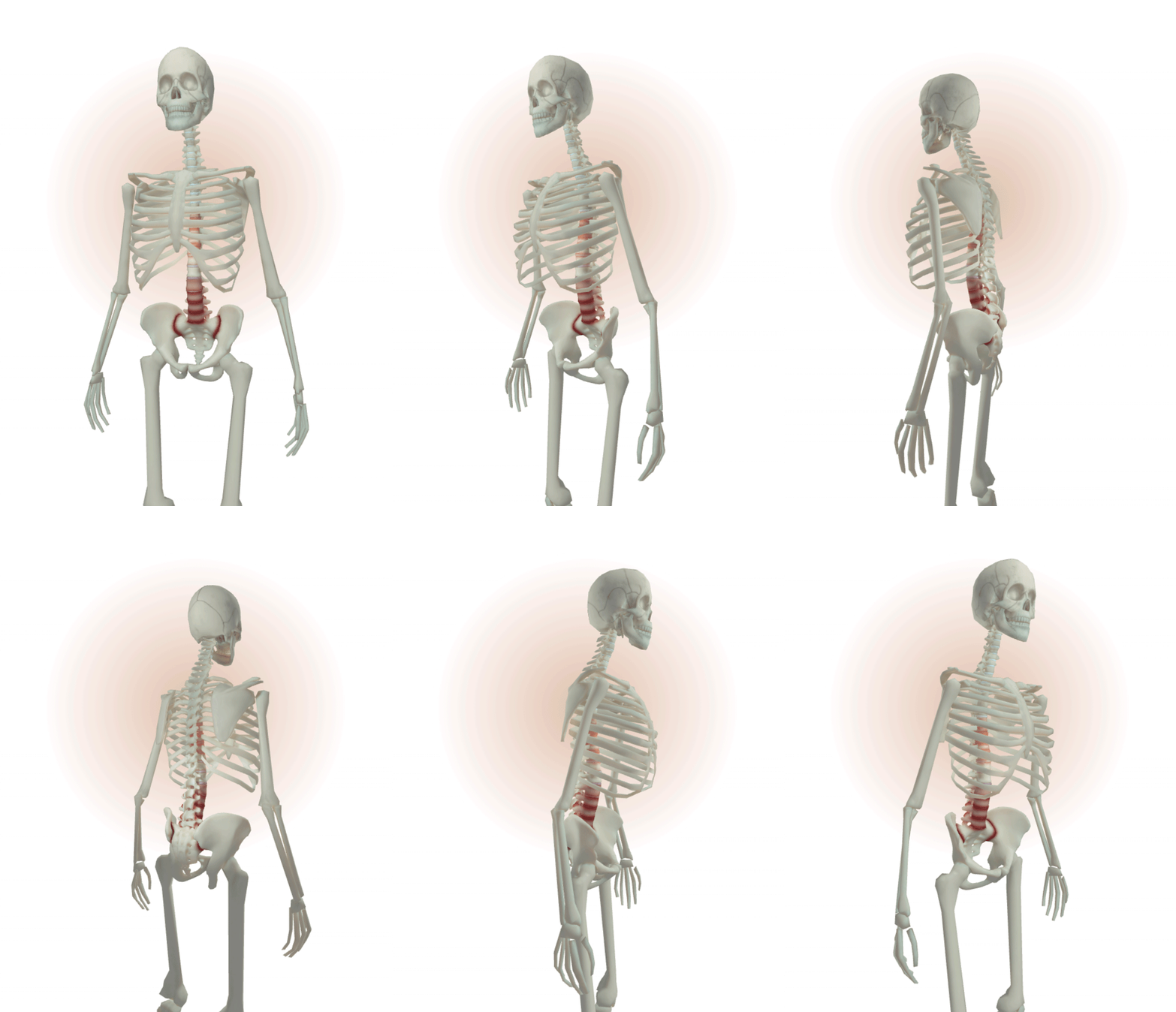
Early stage of inflammation and small areas of erosion in both SIJs and corner of vertebral bodies in the lumbar area (Reference 3)

Models for illustrative purposes only. Not representative of all cases.


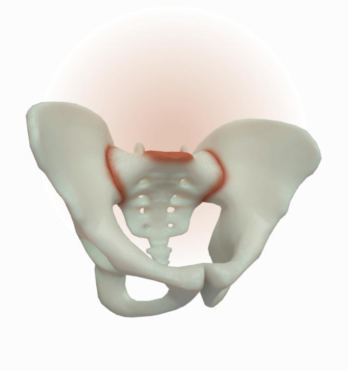
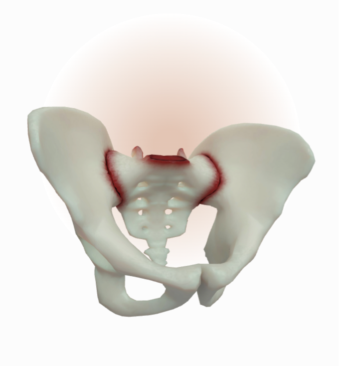
Inflammation and small areas of erosion in both SIJs (Reference 3)

Models for illustrative purposes only. Not representative of all cases.

SIJ Skeletal Changes

Early abnormalities in AS/r-axSpA usually include inflammation and small localized areas of erosion in the SIJs1-4

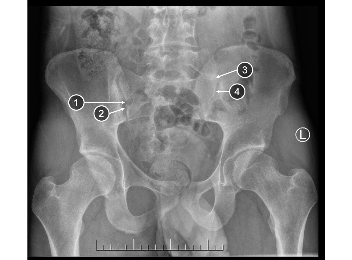
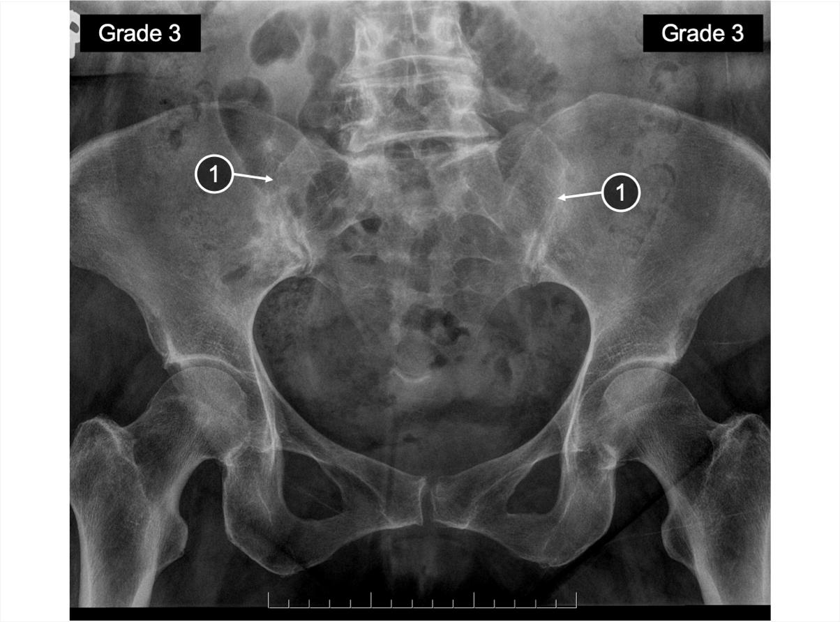
Early | SIJ X-ray

An X-ray of SIJ showing erosion and changes in joint space
The SIJs show widening and narrowing of the joint space, and an erosion in the right SIJ, all of which are consistent with Grade 3 sacroiliitis3

  1. Erosion
  2. Joint space widening
  3. Joint space narrowing
  4. Irregular joint space

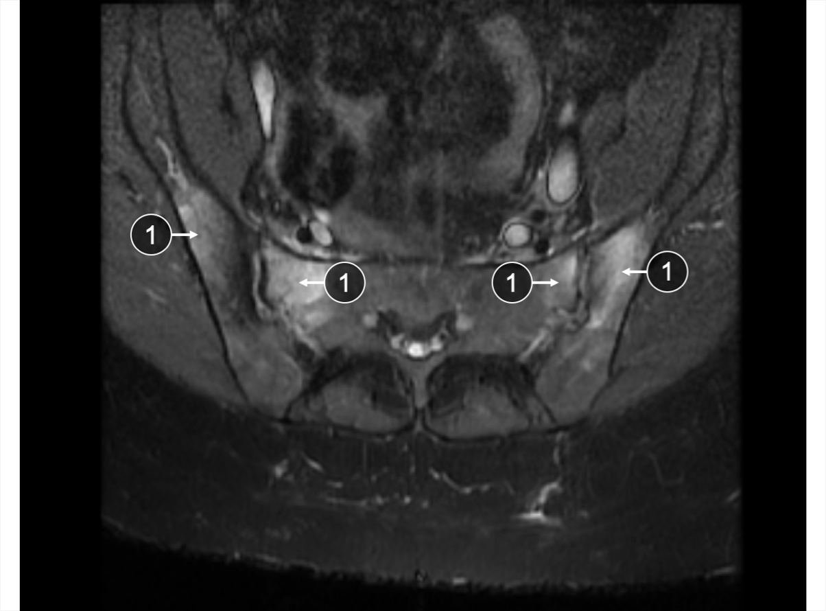
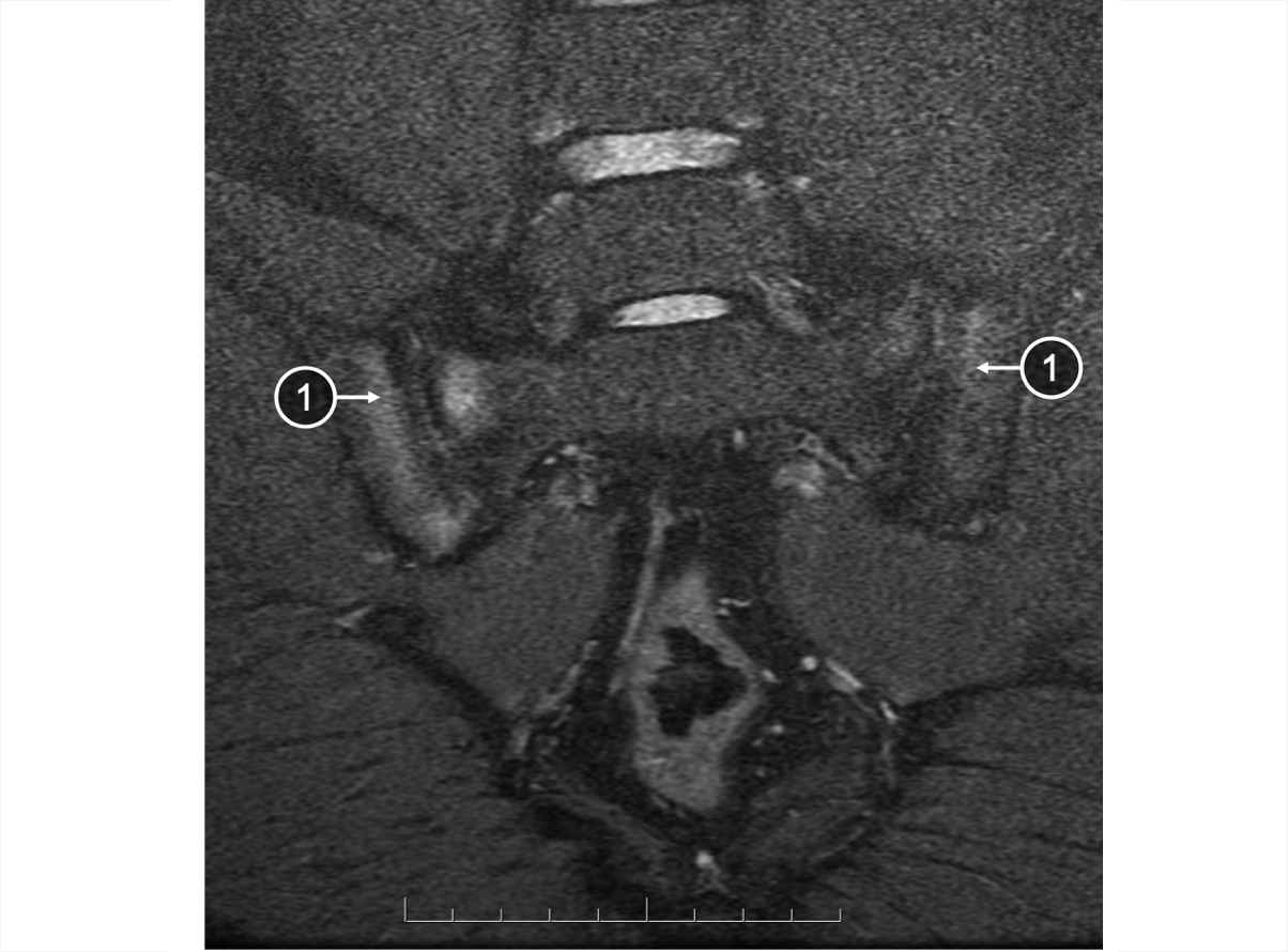
Early | SIJ MRI

An MRI of SIJ showing bone marrow edema
Bone marrow edema, indicative of inflammation, in both SIJs is detected using STIR sequence1,3,5

  1. Bone marrow edema

Images courtesy of Dr. R. Inman (2019) with permission to use in the AS/r-axSpA 3D Explorer Tool for educating HCPs globally.


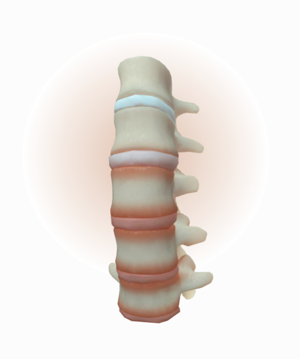
Inflammation at the corner of 4 vertebral bodies in the lumbar area

Models for illustrative purposes only. Not representative of all cases.

Spine Skeletal Changes

Inflammation in the lumbar area also occurs early in AS/r-axSpA5,6


Early | Spine X-ray

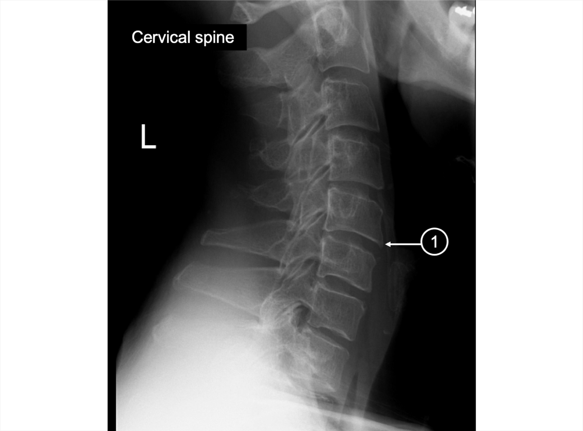
An X-ray of the cervical spine showing small osteophyte
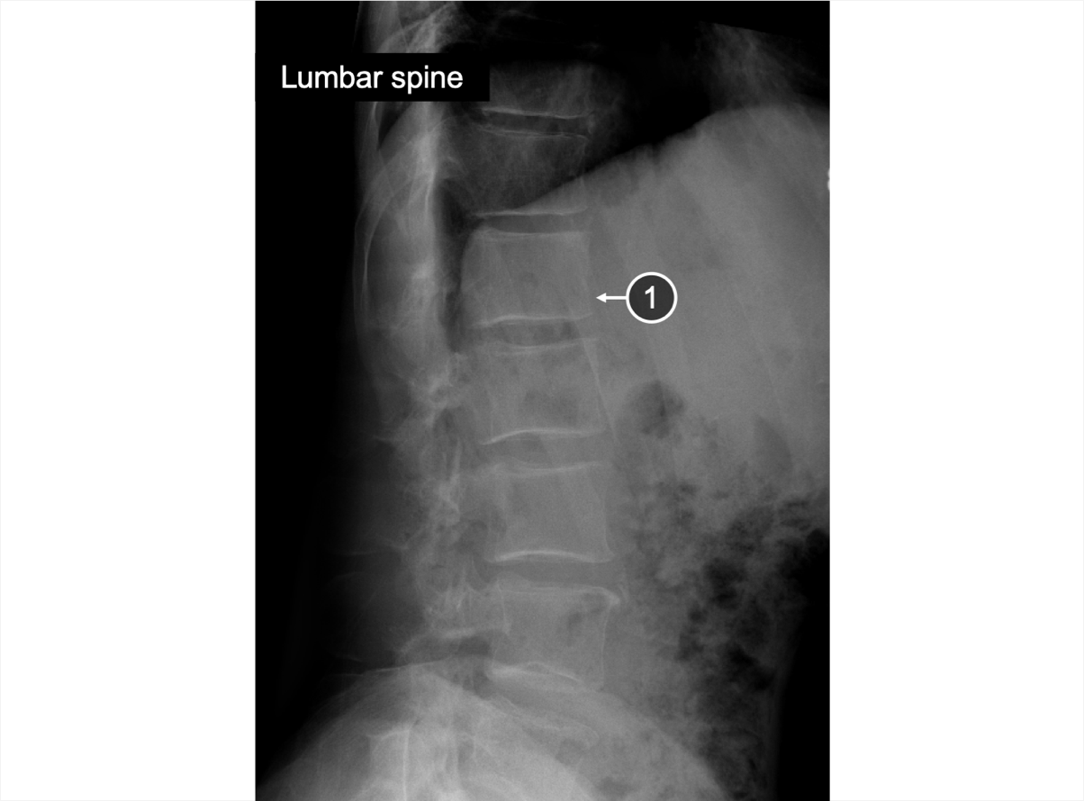
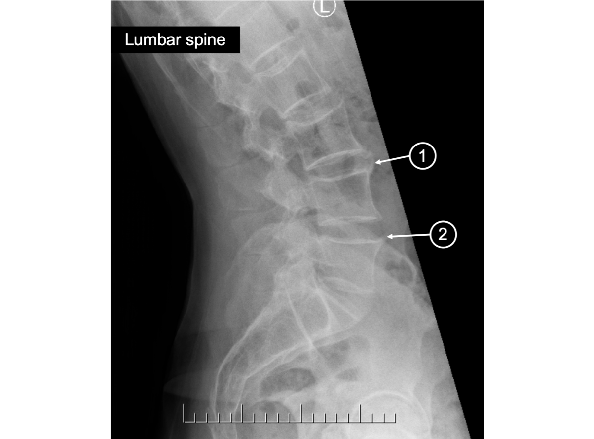
An X-ray of the lumbar spine showing squaring
An X-ray of the cervical spine showing small osteophyte
An X-ray of the lumbar spine showing squaring
Some squaring can be seen on X-ray of the lower lumbar spine3

  1. Squaring

Images courtesy of Dr. R. Inman (2019) with permission to use in the AS/r-axSpA 3D Explorer Tool for educating HCPs globally.

Early | Spine MRI

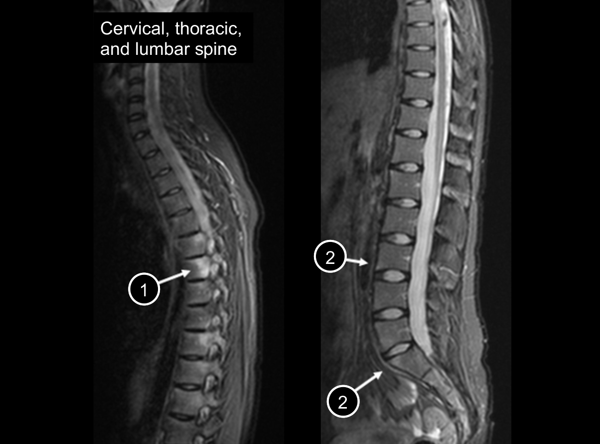
An MRI of the cervical and lumbar spine bone marrow edema
STIR images of the whole spine show signs of disease activity in thoracic and lumbar vertebral bodies5

These are typical sites affected in AS/r-axSpA7

  1. Bone marrow edema in vertebral bodies (posterior aspect)
  2. Bone marrow edema at the corner of vertebral bodies (anterior aspect)

Image reproduced with permission from SPA-imaging.org (www.spa-imaging.org).5


Pathophysiological Changes

  • Inflammation, typically starting at the SIJs, is usually the first pathogenic mechanism in AS/r-axSpA8

  • Enthesitis also occurs from early stages of the disease and is usually axial, although some patients may present with peripheral enthesitis at early stages as well4,9

  • In the spine, one of the early radiological signs is visualized at the corners of the vertebral bodies and is sometimes called "the shiny corner sign"3,4
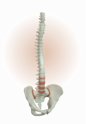
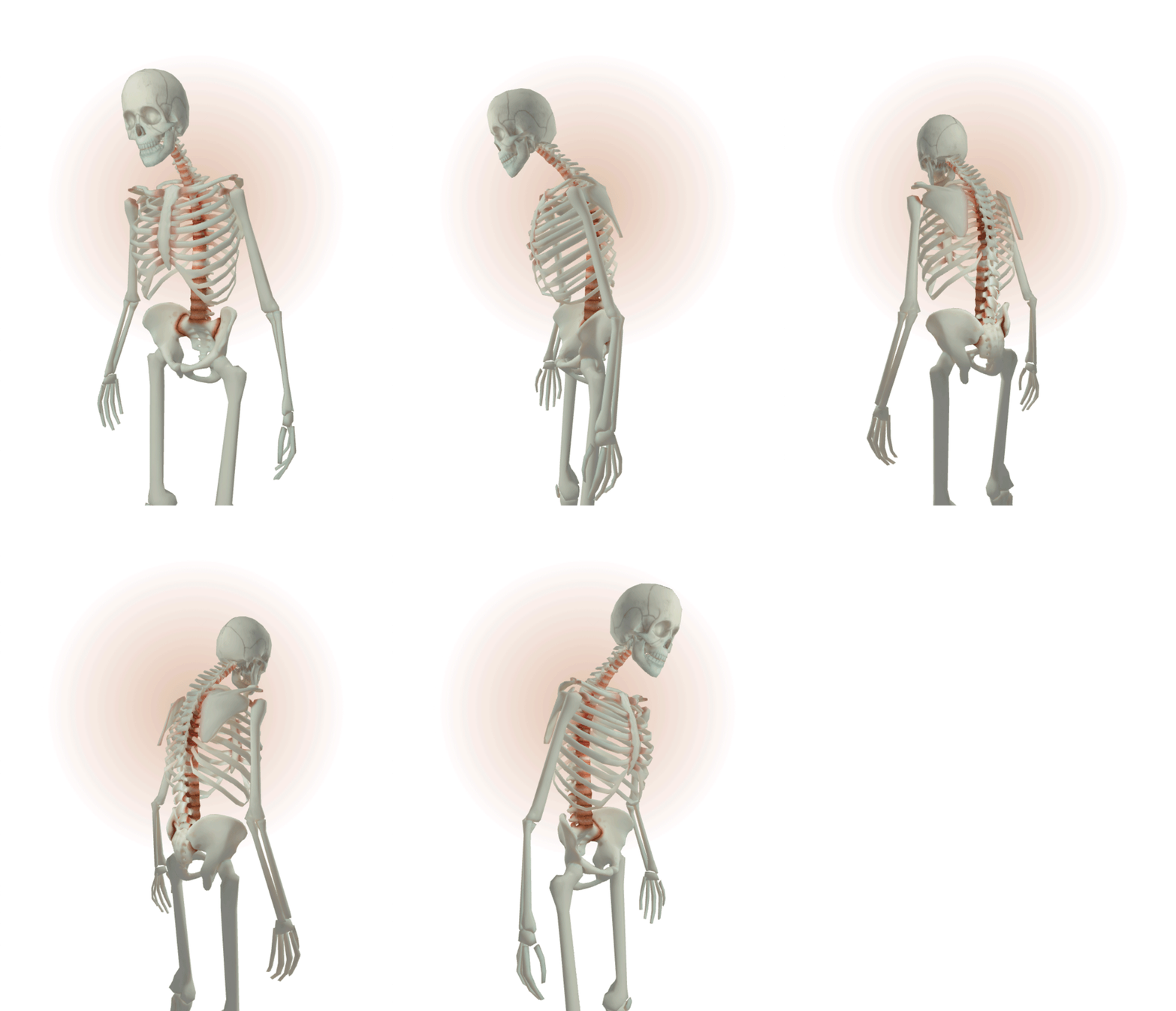
Early stage includes inflammation of both SIJ and spine causing pathological changes

Models for illustrative purposes only. Not representative of all cases.

Patient symptoms

As the disease persists, patients with AS/r-axSpA may present with23,24,25 :

  • Middle back pain of much longer duration, usually 8 to 10 years

  • Difficulty bending over

  • Morning stiffness lasting 2 to 3 hours

  • Profound fatigue

Inflammation, erosion, and some narrowing/widening of joint spaces on SIJs and spinal segments with syndesmophytes (Reference 11)

Models for illustrative purposes only. Not representative of all cases.


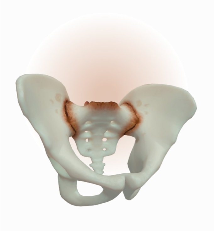
Inflammation, erosion, and some narrowing/widening of joint spaces on both sides of SIJ

Models for illustrative purposes only. Not representative of all cases.

SIJ Skeletal Changes

Inflammation, evidence of progressive erosions, and joint space alteration with areas of widening and/or narrowing are characteristic findings in mid-stage AS/r-axSpA3,10

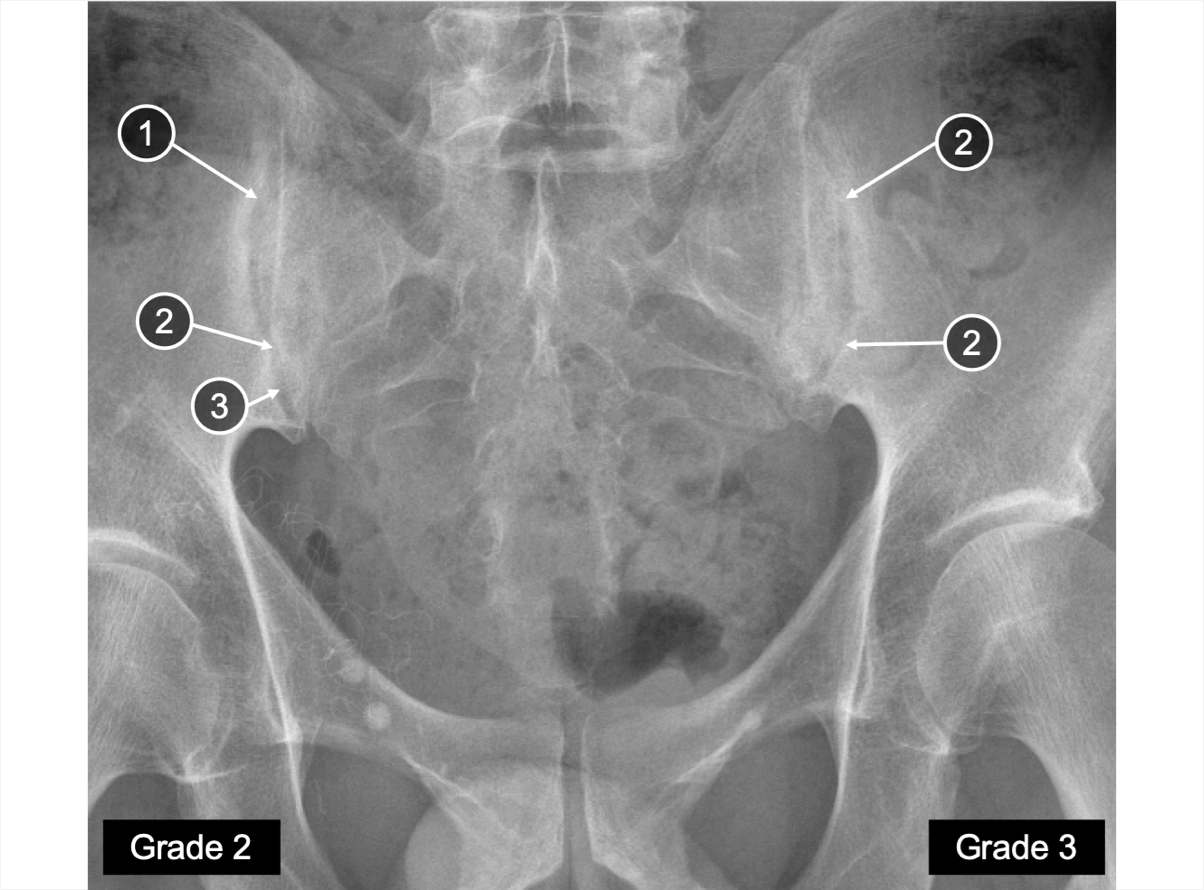
Mid | SIJ X-ray

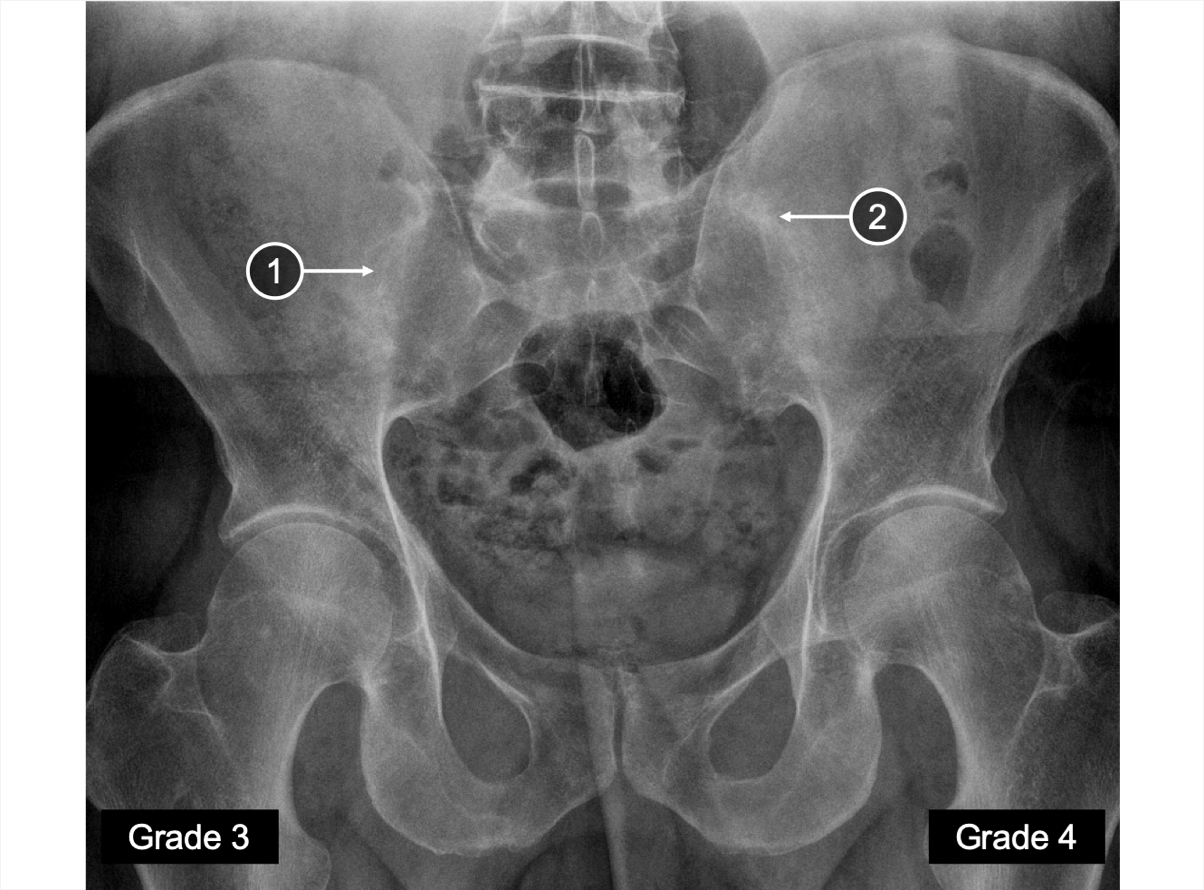
An X-ray of SIJ showing areas of ankylosis
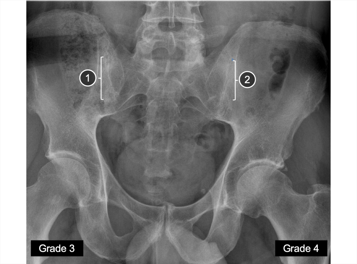
An X-ray of SIJ showing erosion and some narrowing/widening of joint space
An X-ray of SIJ showing areas of ankylosis
An X-ray of SIJ showing erosion and some narrowing/widening of joint space
Grade 2 sacroiliitis defined by inflammation and localized areas of erosion is seen on one side, and Grade 3 sacroiliitis that involves progressive erosion, and widening and narrowing of joint space is seen on the other side3

  1. Irregular joint space with areas of widening
  2. Joint space with severe narrowing
  3. Erosion

Mid | SIJ MRI

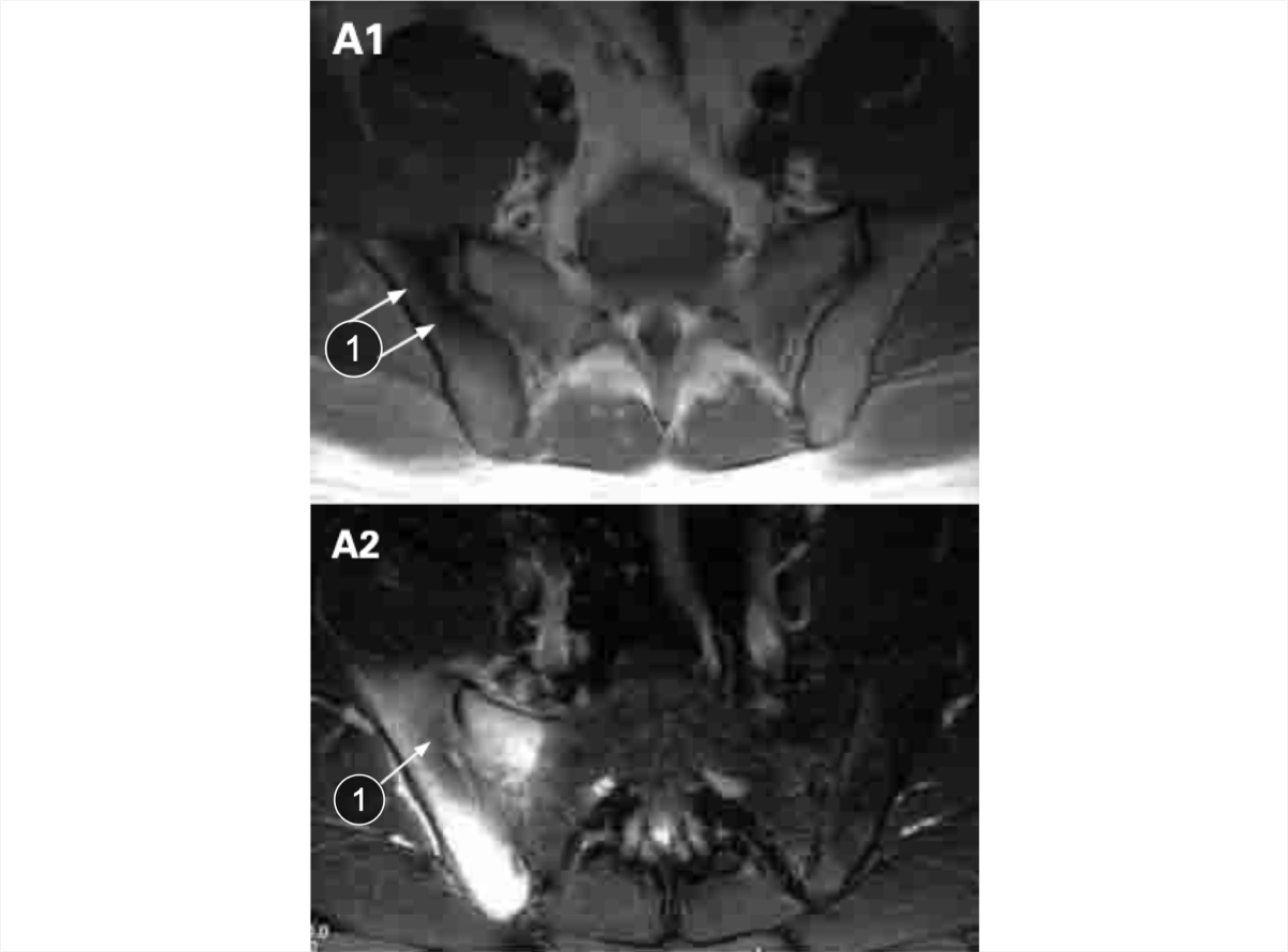
An MRI of SIJ showing bone marrow edema
Bone marrow edema in both SIJs is observed on MRI STIR sequence3,10

  1. Bone marrow edema

Images courtesy of Dr. R. Inman (2019) with permission to use in the AS/r-axSpA 3D Explorer Tool for educating HCPs globally.


Inflammation in lumbar and thoracic spinal segments - with erosion, syndesmophytes, and bridging of joints (Reference 11)

Models for illustrative purposes only. Not representative of all cases.

Spine Skeletal Changes

In addition to inflammation, abnormalities in mid-stage AS/r-axSpA can involve syndesmophytes and some fusion of facet joints3,10


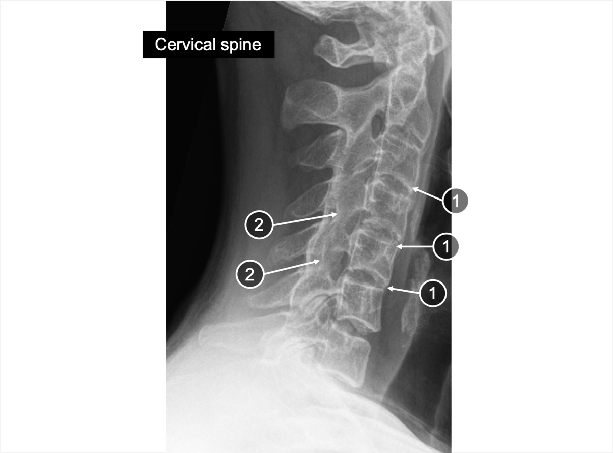
Mid | Spine X-ray

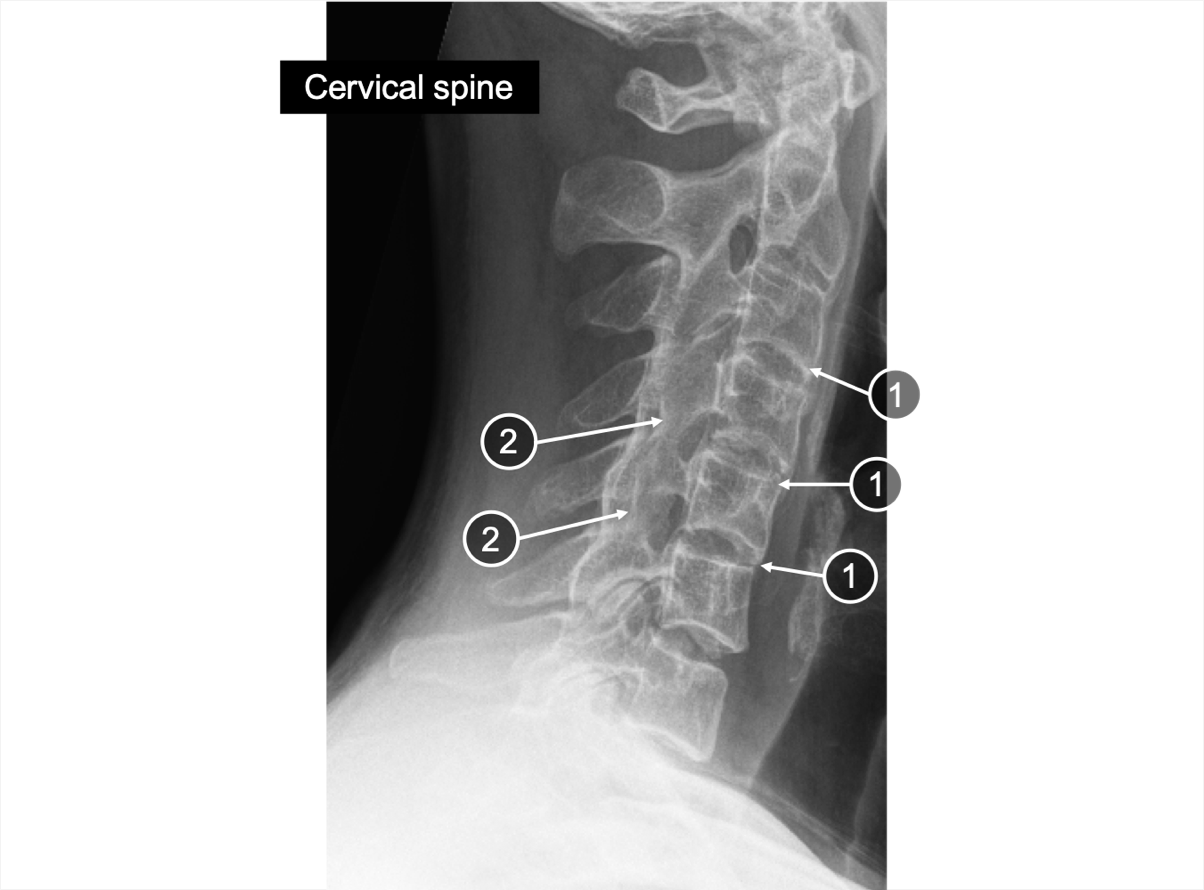
An X-ray of the cervical spine showing erosion and syndesmophyte
Erosion combined with syndesmophytes in patients with AS/r-axSpA are indicative of a more advanced stage of disease5,11

  1. Erosion
  2. Syndesmophyte

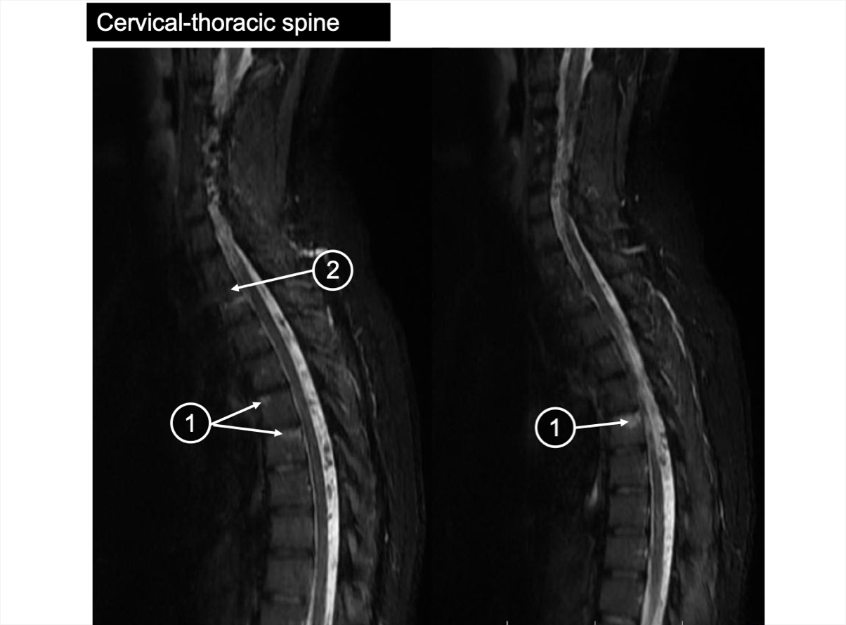
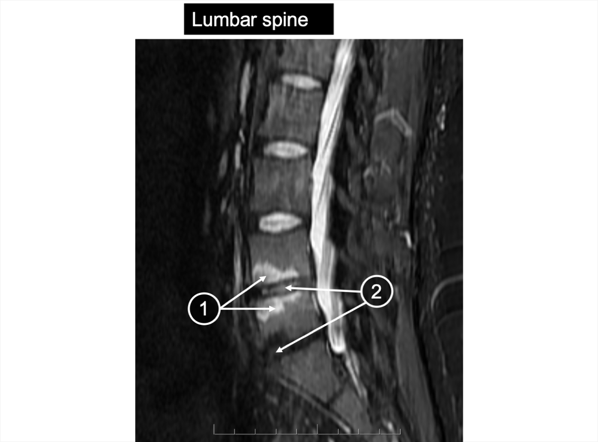
Mid | Spine MRI

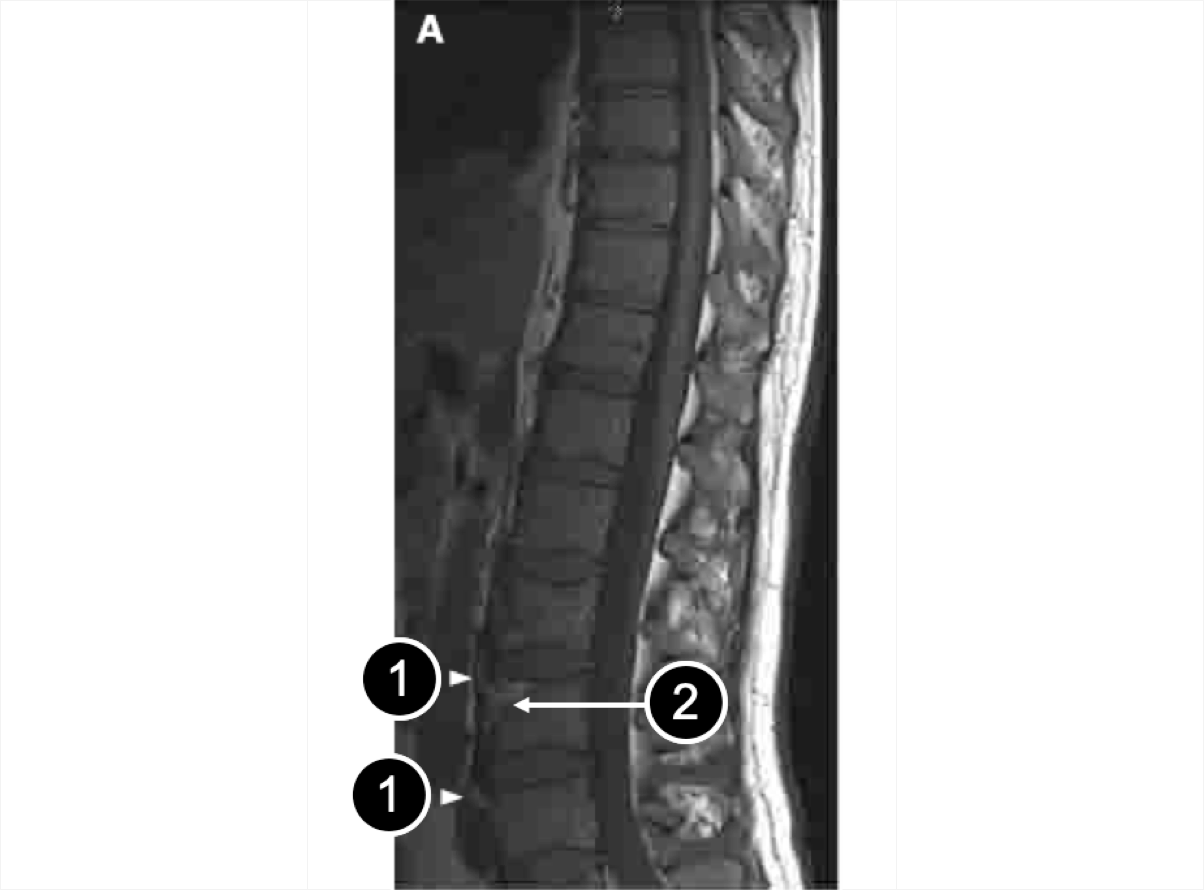
An MRI of the cervical-thoracic spine showing bone marrow edema and discitis
An MRI of the lumbar spine showing bone marrow edema and discitis
An MRI of the cervical-thoracic spine showing bone marrow edema and discitis
An MRI of the lumbar spine showing bone marrow edema and discitis
Bone marrow edema in vertebral bodies and spondylodiscitis, both indicative of active inflammation, can be detected by STIR sequence3,5

  1. Bone marrow edema
  2. Discitis

Images courtesy of Dr. R. Inman (2019) with permission to use in the AS/r-axSpA 3D Explorer Tool for educating HCPs globally.


Pathophysiological Changes

  • Ongoing inflammation in AS/r-axSpA contributes to increasingly pronounced bone erosions24,25

  • Inflammation is followed by a repair process that triggers pathogenic bone formation12,13
Mid stage has inflammation, erosion, syndesmophytes, some joint spaces changes on SIJs and spine causing pathological changes

Models for illustrative purposes only. Not representative of all cases.

Patient symptoms

In the late stage of the disease, patients with AS/r-axSpA may present with23,24,25 :

  • Neck and middle back pain in addition to lower back pain that has been ongoing for a long time, usually 20 years or more

  • Difficulty performing everyday activities such as tying shoelaces and checking blind spots

  • Mild shortness of breath with exertion

  • Fatigue

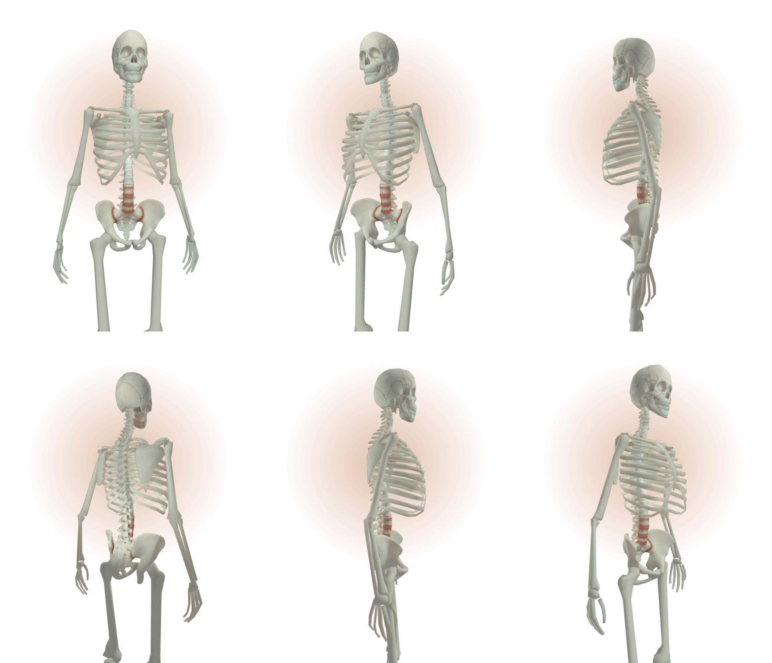
Inflammation is present, loss of joint space with ankylosing in SIJs, vertebrae affected along the spine with fused joints (Reference 11)

Models for illustrative purposes only. Not representative of all cases.


Inflammation is still present with loss of joint space and ankylosing in SIJs

Models for illustrative purposes only. Not representative of all cases.

SIJ Skeletal Changes

Typical SIJ changes at this stage of AS/r-axSpA include partial or complete bone fusion (ankylosis)3,10

Late | SIJ X-ray

An X-ray of the SIJs showing different degree of fusion
X-rays indicate partial or complete fusion of the SIJs (ankylosis)3

  1. Almost complete fusion
  2. Complete fusion

Images courtesy of Dr. R. Inman (2019) with permission to use in the AS/r-axSpA 3D Explorer Tool for educating HCPs globally.

Late | SIJ MRI

An MRI of the SIJs showing areas of sclerosis
MRIs reveal areas of sclerosis3

  1. Areas of sclerosis

Images reprinted with permission. © 2009. BMJ. All rights reserved.


Inflammation is present, vertebrae affected throughout the spine and fused facet joints present in each segment (Reference 11)

Models for illustrative purposes only. Not representative of all cases.

Spine Skeletal Changes

Late-stage disease can be characterized by bridging syndesmophytes and fused facet joints affecting several segments of the spine3,10


Late | Spine X-ray

An X-ray of the cervical spine showing briding of syndesmophyte and fused facet joints
An X-ray of the lumbar spine showing briding of syndesmophyte
An X-ray of the cervical spine showing briding of syndesmophyte and fused facet joints
An X-ray of the lumbar spine showing briding of syndesmophyte
The X-ray images illustrate bridging syndesmophytes and fused facet joints in multiple segments of the spine, which are indicative of advanced AS/r-axSpA3,5
  1. Bridging syndesmophyte

Images courtesy of Dr. R. Inman (2019) with permission to use in the AS/r-axSpA 3D Explorer Tool for educating HCPs globally.

Late | Spine MRI

An MRI of the thoracic- lumbar spine showing syndesmophyte and area of inflammation
Similar to the X-ray images, the MRI reveals bridging syndesmophytes5

At this stage, inflammation is still present12

  1. Syndesmophyte
  2. Area of inflammation

Images reprinted with permission. © 2009. BMJ. All rights reserved.3


Pathophysiological Changes

  • At this stage, pathogenic bone formation leads to progressive spinal fusion13

  • This can contribute to the rigidity of the spine, causing an increase in physical limitations for patients13
Late stage has inflammation, loss of joint space with ankylosing in SIJs, & fused joints in spine causing pathological change

Models for illustrative purposes only. Not representative of all cases.

References

  1. Van Mechelen M, Gulino GR, de Valm K, Lories R. Bone disease in axial spondyloarthritis. Calcif Tissue Int. 2018;102(5):547-558.
  2. Maksymowych WP, Inman RD, Salonen D, et al. Spondyloarthritis Research Consortium of Canada magnetic resonance imaging index for assessment of sacroiliac joint inflammation in ankylosing spondylitis. Arthritis Rheum. 2005;53(5):703-709.
  3. Sieper J, Rudwaleit M, Baraliakos X, et al. The Assessment of SpondyloArthritis international Society (ASAS) handbook: a guide to assess spondyloarthritis. Ann Rheum Dis. 2009;68(Suppl 2):ii1-ii44.
  4. Østergaard M, Lambert RGW. Imaging in ankylosing spondylitis. Ther Adv Musculoskel Dis. 2012;4(4):301-311.
  5. Jurik, A. Spondylarthropathy Imaging. www.spa-imaging.org. Accessed July 26, 2023.
  6. Neerinckx B, Lories RJ. Structural disease progression in axial spondyloarthritis: still a cause for concern? Curr Rheumatol Rep. 2017;19:14.
  7. Maksymowych WP, Inman RD, Salonen D, et al. Spondyloarthritis Research Consortium of Canada magnetic resonance imaging index for assessment of spinal inflammation in ankylosing spondylitis. Arthritis Rheum. 2005;53(4):502-509
  8. Raychaudhuri SK, Saxena A, Raychaudhuri SP. Role of IL-17 in the pathogenesis of psoriatic arthritis and axial spondyloarthritis. Clin Rheumatol. 2015;34(6):1019-1023.
  9. Watad A, Bridgewood C, Russell T, et al. The early phases of ankylosing spondylitis: emerging insights from clinical and basic science. Front Immunol. 2018;9:2668.
  10. Pialat JB, Di Marco L, Feydy A, et al. Sacroiliac joints imaging in axial spondyloarthritis. Diagn Interv Imaging. 2016;97(7-8):697-708.
  11. Braun J, van der Heijde D, Dougados M, et al. Staging of patients with ankylosing spondylitis: a preliminary proposal. Ann Rheum Dis. 2002;61(Suppl III):iii19-iii23.
  12. Poddubnyy D, Sieper J. Mechanism of new bone formation in axial spondyloarthritis. Curr Rheumatol Rep. 2017;19(9):55.
  13. Osta B, Benedetti G, Miossec P. Classical and paradoxical effects of TNF-α on bone homeostasis. Front Immunol. 2014;5:48.
  14. Baraliakos X, Braun J. Non-radiographic axial spondyloarthritis and ankylosing spondylitis: what are the similarities and differences? RMD Open. 2015;1(Suppl 1):e000053.
  15. Kiltz U, Baraliakos X, Regel A, et al. Causes of pain in patients with axial spondyloarthritis. Clin Exp Rheumatol. 2017;35(Suppl 107):S102-107.
  16. Deodhar A, Reveille JD, van den Bosch F, et al. The concept of axial spondyloarthritis: joint statement of the spondyloarthritis research and treatment network and the Assessment of SpondyloArthritis international Society in response to the US Food and Drug Administration's comments and concerns. Arthritis & Rheumatol. 2014;66(10):2649-2656.
  17. Bubová K, Forejtová Š, Zegzulková K, et al. Cross-sectional study of patients with axial spondyloarthritis fulfilling imaging arm of ASAS classification criteria: baseline clinical characteristics and subset differences in a single-centre cohort. BMJ Open. 2019;9:e024713.
  18. Ghosh N, Ruderman EM. Nonradiographic axial spondyloarthritis: clinical and therapeutic relevance. Arthritis Res Ther. 2017;19(1):286.
  19. Deodhar A, van der Heijde D, Gensler LS, et al. Ixekizumab for patients with non-radiographic axial spondyloarthritis (COAST-X): a randomised, placebo-controlled trial. Lancet. 2020;395(10217):53-64.
  20. Rudwaleit M, Landewé R, van der Heijde D, et al. The development of Assessment of SpondyloArthritis international Society classification criteria for axial spondyloarthritis (part 1): classification of paper patients by expert opinion including uncertainty appraisal. Ann Rheum Dis. 2009;68(6):770-776.
  21. Poddubnyy D, Rudwaleit M, Haibel H, et al. Rates and predictors of radiographic sacroiliitis progression over 2 years in patients with axial spondyloarthritis. Ann Rheum Dis. 2011;70(8):1369-1374.
  22. Zhang S, Wang Y, Peng L, et al. Comparison of clinical features in HLA-B27 positive and negative patients with axial spondyloarthritis: results from a cohort of 4,131 patients. Front Med (Lausanne). 2020;7:609562.
  23. Barhum, L. Verywell Health. www.verywellhealth.com/axial-spondyloarthritis-progression-6890574. Accessed July 26, 2023.
  24. Danve, A., Deodhar, A. Treatment of axial spondyloarthritis: an update. Nat Rev Rheumatol 18, 205–216 (2022). https://doi.org/10.1038/s41584-022-00761-z
  25. Schwartzman, S., Ruderman, EMR. A Road Map of the Axial Spondyloarthritis Continuum. Mayo Clin Proc. 2022;97(1):134-145

Abbreviations

AS: ankylosing spondylitis; ASAS: Assessment of SpondyloArthritis international Society; CRP: C-reactive protein; HCP: health care practitioner; HLA-B27: human leukocyte antigen B27; mNY: modified New York; MRI: magnetic resonance imaging; nr-axSpA: nonradiographic axial spondyloarthritis; NSAID: nonsteroidal anti-inflammatory drug; r-axSpA: radiographic axial spondyloarthritis; SIJ: sacroiliac joint; SpA: spondyloarthritis; STIR: short tau inversion recovery


VV-MED-141368

Please rate your satisfaction with the content on the following statements:

Very Dissatisfied

Dissatisfied

Neutral

Satisfied

Very Satisfied
Credibility of information
Relevance of the information to my needs
Confidence to implement the learnings in my clinical practice

Please rate your satisfaction with the content on the following statements:

Credibility of information
Very Satisfied
Very Dissatisfied
Relevance of the information to my needs
Very Satisfied
Very Dissatisfied
Confidence to implement the learnings in my clinical practice
Very Satisfied
Very Dissatisfied
  • Copyright
  • Terms of Use
  • Privacy Statement
  • Consumer Health Privacy Notice
  • Accessibility Statement
  • Sitemap

To speak to customer support:
Call (XXX) XXX-XXXX

This site is intended for US Healthcare Professionals only.

4.4.53 05/2025 | GLOOTH00001 04/2015 | © Lilly USA, LLC 2025. All rights reserved.

Product names listed above are trademarks or registered trademarks owned by or licensed to Eli Lilly and Company, its subsidiaries, or affiliates

California Consumer Privacy Act (CCPA) Opt-Out Icon Your Privacy Choices
Cookie Settings
Facebook X LinkedIn
Lilly